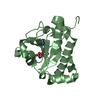
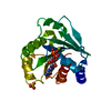
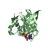
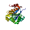
- PDB-1fzq: CRYSTAL STRUCTURE OF MURINE ARL3-GDP -
+
Open data
ID or keywords:
Loading...
-
Basic information
Entry
Database: PDB / ID: 1fzq
Title
CRYSTAL STRUCTURE OF MURINE ARL3-GDP
Components
ADP-RIBOSYLATION FACTOR-LIKE PROTEIN 3
Keywords
SIGNALING PROTEIN / Protein-GDP complex without magnesium / ARF family / Ras Superfamily / G-domain
Function / homology
Function and homology information
Trafficking of myristoylated proteins to the cilium / photoreceptor cell development / protein localization to ciliary membrane / intraciliary transport / post-Golgi vesicle-mediated transport / photoreceptor connecting cilium / Golgi to plasma membrane transport / protein localization to cilium / smoothened signaling pathway / small GTPase-mediated signal transduction ...Trafficking of myristoylated proteins to the cilium / photoreceptor cell development / protein localization to ciliary membrane / intraciliary transport / post-Golgi vesicle-mediated transport / photoreceptor connecting cilium / Golgi to plasma membrane transport / protein localization to cilium / smoothened signaling pathway / small GTPase-mediated signal transduction / ciliary transition zone / mitotic cytokinesis / cilium assembly / axoneme / cytoplasmic microtubule / spindle microtubule / kidney development / GDP binding / protein transport / microtubule cytoskeleton / midbody / microtubule binding / cilium / ciliary basal body / Golgi membrane / GTPase activity / centrosome / GTP binding / magnesium ion binding / Golgi apparatus / nucleoplasm / nucleus / cytoplasm Similarity search - Function
ADP-ribosylation factor-like protein 2/3 / Small GTPase Arf domain profile. / Sar1p-like members of the Ras-family of small GTPases / Small GTPase superfamily, ARF/SAR type / ADP-ribosylation factor family / ARF-like small GTPases; ARF, ADP-ribosylation factor / Rab subfamily of small GTPases / Small GTP-binding protein domain / P-loop containing nucleotide triphosphate hydrolases / Rossmann fold ...ADP-ribosylation factor-like protein 2/3 / Small GTPase Arf domain profile. / Sar1p-like members of the Ras-family of small GTPases / Small GTPase superfamily, ARF/SAR type / ADP-ribosylation factor family / ARF-like small GTPases; ARF, ADP-ribosylation factor / Rab subfamily of small GTPases / Small GTP-binding protein domain / P-loop containing nucleotide triphosphate hydrolases / Rossmann fold / P-loop containing nucleoside triphosphate hydrolase / 3-Layer(aba) Sandwich / Alpha Beta Similarity search - Domain/homology
The biological active unit is most probably a monomer. The electron density map showed a disulphide bridge between Cys 118 and a symmetry-related Cys 118 of a crystal neighbour. This is considered to be a crystallization artefact and was not included in the model.
-
Components
-
Protein , 1 types, 1 molecules A
#1: Protein
ADP-RIBOSYLATIONFACTOR-LIKEPROTEIN3 / ARF-LIKE PROTEIN 3 / ARL3
Mass: 20380.307 Da / Num. of mol.: 1 Source method: isolated from a genetically manipulated source Source: (gene. exp.) Mus musculus (house mouse) / Plasmid: PET21D / Production host: Escherichia coli (E. coli) / References: UniProt: Q9WUL7
In the structure databanks used in Yorodumi, some data are registered as the other names, "COVID-19 virus" and "2019-nCoV". Here are the details of the virus and the list of structure data.
Jan 31, 2019. EMDB accession codes are about to change! (news from PDBe EMDB page)
EMDB accession codes are about to change! (news from PDBe EMDB page)
The allocation of 4 digits for EMDB accession codes will soon come to an end. Whilst these codes will remain in use, new EMDB accession codes will include an additional digit and will expand incrementally as the available range of codes is exhausted. The current 4-digit format prefixed with “EMD-” (i.e. EMD-XXXX) will advance to a 5-digit format (i.e. EMD-XXXXX), and so on. It is currently estimated that the 4-digit codes will be depleted around Spring 2019, at which point the 5-digit format will come into force.
The EM Navigator/Yorodumi systems omit the EMD- prefix.
Related info.:Q: What is EMD? / ID/Accession-code notation in Yorodumi/EM Navigator
Yorodumi is a browser for structure data from EMDB, PDB, SASBDB, etc.
This page is also the successor to EM Navigator detail page, and also detail information page/front-end page for Omokage search.
The word "yorodu" (or yorozu) is an old Japanese word meaning "ten thousand". "mi" (miru) is to see.
Related info.:EMDB / PDB / SASBDB / Comparison of 3 databanks / Yorodumi Search / Aug 31, 2016. New EM Navigator & Yorodumi / Yorodumi Papers / Jmol/JSmol / Function and homology information / Changes in new EM Navigator and Yorodumi
 Movie
Movie Controller


 Open data
 Basic information
 Components
 Keywords
 Function and homology information
 X-RAY DIFFRACTION /
  Authors
 Citation
 Structure visualization
 Downloads & links
 Other downloads
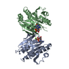


 PDBj
PDBj
 Assembly


 Mass: 96.063 Da / Num. of mol.: 2 / Source method: obtained synthetically / Formula: SO4
 Mass: 18.038 Da / Num. of mol.: 1 / Source method: obtained synthetically / Formula: H4N
 Mass: 195.237 Da / Num. of mol.: 1 / Source method: obtained synthetically / Formula: C6H13NO4S / Comment: pH buffer*YM
 Type: RNA linking / Mass: 443.201 Da / Num. of mol.: 1 / Source method: obtained synthetically / Formula: C10H15N5O11P2 / Comment: GDP, energy-carrying molecule*YM
 Sample preparation
 / Beamline: ID13 / Wavelength: 0.782
  Processing