 Movie
Movie Controller

[English] 日本語
 Yorodumi
- PDB-1fzd: STRUCTURE OF RECOMBINANT ALPHAEC DOMAIN FROM HUMAN FIBRINOGEN-420 -
+ Open data

- Basic information
| Entry | Database: PDB / ID: 1fzd | |||||||||
|---|---|---|---|---|---|---|---|---|---|---|
| Title | STRUCTURE OF RECOMBINANT ALPHAEC DOMAIN FROM HUMAN FIBRINOGEN-420 | |||||||||
 Components Components | FIBRINOGEN-420 | |||||||||
 Keywords Keywords | BLOOD COAGULATION / FIBRINOGEN-420 / ALPHAEC DOMAIN / FIBRINOGEN RELATED DOMAIN / GLYCOSYLATED PROTEIN | |||||||||
| Function / homology |  Function and homology information Function and homology informationblood coagulation, common pathway / induction of bacterial agglutination / fibrinogen complex / platelet alpha granule / Regulation of TLR by endogenous ligand / MyD88 deficiency (TLR2/4) / positive regulation of heterotypic cell-cell adhesion / IRAK4 deficiency (TLR2/4) / MyD88:MAL(TIRAP) cascade initiated on plasma membrane / plasminogen activation ...blood coagulation, common pathway / induction of bacterial agglutination / fibrinogen complex / platelet alpha granule / Regulation of TLR by endogenous ligand / MyD88 deficiency (TLR2/4) / positive regulation of heterotypic cell-cell adhesion / IRAK4 deficiency (TLR2/4) / MyD88:MAL(TIRAP) cascade initiated on plasma membrane / plasminogen activation / extracellular matrix structural constituent / p130Cas linkage to MAPK signaling for integrins / positive regulation of peptide hormone secretion / GRB2:SOS provides linkage to MAPK signaling for Integrins / positive regulation of exocytosis / blood coagulation, fibrin clot formation / protein polymerization / negative regulation of endothelial cell apoptotic process / positive regulation of vasoconstriction / Integrin cell surface interactions / : / negative regulation of extrinsic apoptotic signaling pathway via death domain receptors / fibrinolysis / Integrin signaling / positive regulation of substrate adhesion-dependent cell spreading / platelet alpha granule lumen / cell-matrix adhesion / positive regulation of protein secretion / Post-translational protein phosphorylation / Signaling by high-kinase activity BRAF mutants / MAP2K and MAPK activation / response to calcium ion / platelet aggregation / Regulation of Insulin-like Growth Factor (IGF) transport and uptake by Insulin-like Growth Factor Binding Proteins (IGFBPs) / Signaling by RAF1 mutants / Signaling by moderate kinase activity BRAF mutants / Paradoxical activation of RAF signaling by kinase inactive BRAF / Signaling downstream of RAS mutants / Signaling by BRAF and RAF1 fusions / Platelet degranulation / extracellular vesicle / extracellular matrix / ER-Phagosome pathway / protein-containing complex assembly / blood microparticle / adaptive immune response / positive regulation of ERK1 and ERK2 cascade / protein-macromolecule adaptor activity / endoplasmic reticulum lumen / Amyloid fiber formation / external side of plasma membrane / signaling receptor binding / innate immune response / structural molecule activity / cell surface / endoplasmic reticulum / : / extracellular exosome / extracellular region / metal ion binding / plasma membrane Similarity search - Function | |||||||||
| Biological species |  Homo sapiens (human) Homo sapiens (human) | |||||||||
| Method |  X-RAY DIFFRACTION / SYNCHROTRON / MOLECULAR REPLACEMENT / Resolution: 2.1 Å X-RAY DIFFRACTION / SYNCHROTRON / MOLECULAR REPLACEMENT / Resolution: 2.1 Å | |||||||||
 Authors Authors | Spraggon, G. / Applegate, D. / Everse, S.J. / Zhang, J.-Z. / Veerapandian, L. / Redman, C. / Doolittle, R.F. / Grieninger, G. | |||||||||
 Citation Citation | Journal: Proc.Natl.Acad.Sci.USA / Year: 1998 Title: Crystal structure of a recombinant alphaEC domain from human fibrinogen-420. Authors: Spraggon, G. / Applegate, D. / Everse, S.J. / Zhang, J.Z. / Veerapandian, L. / Redman, C. / Doolittle, R.F. / Grieninger, G. | |||||||||
| History |
|
- Structure visualization
| Structure viewer | Molecule: MolmilJmol/JSmol |
|---|
- Downloads & links
-Download
| PDBx/mmCIF format | 1fzd.cif.gz | 342.5 KB | Display | PDBx/mmCIF format |
|---|---|---|---|---|
| PDB format | pdb1fzd.ent.gz | 279.6 KB | Display | PDB format |
| PDBx/mmJSON format | 1fzd.json.gz | Tree view | PDBx/mmJSON format | |
| Others |  Other downloads Other downloads |
-Validation report
| Arichive directory | https://data.pdbj.org/pub/pdb/validation_reports/fz/1fzdftp://data.pdbj.org/pub/pdb/validation_reports/fz/1fzd | HTTPS FTP |
|---|
-Related structure data
| Related structure data |  1fczS S: Starting model for refinement |
|---|---|
| Similar structure data |


-Links
 PDBj
PDBj


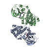
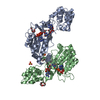
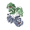
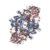
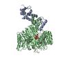
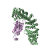
- Assembly
| Deposited unit | 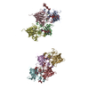
| ||||||||||||||||||||||||||||||||
|---|---|---|---|---|---|---|---|---|---|---|---|---|---|---|---|---|---|---|---|---|---|---|---|---|---|---|---|---|---|---|---|---|---|
| 1 | 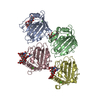
| ||||||||||||||||||||||||||||||||
| 2 | 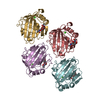
| ||||||||||||||||||||||||||||||||
| Unit cell |
| ||||||||||||||||||||||||||||||||
| Noncrystallographic symmetry (NCS) | NCS oper:
|
-Components
-Protein , 1 types, 8 molecules ABCDEFGH
| #1: Protein | Mass: 22840.660 Da / Num. of mol.: 8 / Fragment: ALPHA-EC DOMAIN Source method: isolated from a genetically manipulated source Source: (gene. exp.) Homo sapiens (human) / Organ: BLOOD / Production host:  Pichia pastoris (fungus) / References: UniProt: P02671 Pichia pastoris (fungus) / References: UniProt: P02671 |
|---|
-Sugars , 4 types, 10 molecules 
| #2: Polysaccharide | Source method: isolated from a genetically manipulated source #3: Polysaccharide | 2-acetamido-2-deoxy-beta-D-glucopyranose-(1-2)-alpha-D-mannopyranose-(1-6)-[alpha-D-mannopyranose- ...2-acetamido-2-deoxy-beta-D-glucopyranose-(1-2)-alpha-D-mannopyranose-(1-6)-[alpha-D-mannopyranose-(1-3)]alpha-D-mannopyranose-(1-4)-2-acetamido-2-deoxy-alpha-D-glucopyranose-(1-4)-2-acetamido-2-deoxy-beta-D-glucopyranose | Source method: isolated from a genetically manipulated source #4: Polysaccharide | Source method: isolated from a genetically manipulated source #6: Sugar | ChemComp-NAG /  Type: D-saccharide, beta linking / Mass: 221.208 Da / Num. of mol.: 4 Type: D-saccharide, beta linking / Mass: 221.208 Da / Num. of mol.: 4Source method: isolated from a genetically manipulated source Formula: C8H15NO6 |
|---|
-Non-polymers , 2 types, 504 molecules 

| #5: Chemical | ChemComp-CA /  Mass: 40.078 Da / Num. of mol.: 8 / Source method: obtained synthetically / Formula: Ca Mass: 40.078 Da / Num. of mol.: 8 / Source method: obtained synthetically / Formula: Ca#7: Water | ChemComp-HOH / | Mass: 18.015 Da / Num. of mol.: 496 / Source method: isolated from a natural source / Formula: H2O |
|---|
-Details
| Has protein modification | Y |
|---|
-Experimental details
-Experiment
| Experiment | Method: X-RAY DIFFRACTION / Number of used crystals: 3 |
|---|
- Sample preparation
| Crystal | Density Matthews: 2.58 Å3/Da / Density % sol: 50 % | ||||||||||||||||||||||||||||||||||||||||||
|---|---|---|---|---|---|---|---|---|---|---|---|---|---|---|---|---|---|---|---|---|---|---|---|---|---|---|---|---|---|---|---|---|---|---|---|---|---|---|---|---|---|---|---|
| Crystal grow | Method: vapor diffusion, sitting drop / pH: 5.5 Details: PROTEIN WAS CRYSTALLIZED BY SITTING DROP VAPOUR DIFFUSION FROM 20% PEG 3350, 0.15 M CACL2, 0.1M IMIDAZOLE- ACETATE PH 5.5 0.002 M SODIUM AZIDE, vapor diffusion - sitting drop | ||||||||||||||||||||||||||||||||||||||||||
| Crystal | *PLUS | ||||||||||||||||||||||||||||||||||||||||||
| Crystal grow | *PLUS pH: 7.5 / Method: vapor diffusion, sitting drop | ||||||||||||||||||||||||||||||||||||||||||
| Components of the solutions | *PLUS
|
-Data collection
| Diffraction | Mean temperature: 293 K |
|---|---|
| Diffraction source | Source: SYNCHROTRON / Site: NSLS  / Beamline: X12C / Wavelength: 1.08 / Beamline: X12C / Wavelength: 1.08 |
| Detector | Detector: CCD / Date: Feb 20, 1998 |
| Radiation | Monochromatic (M) / Laue (L): M / Scattering type: x-ray |
| Radiation wavelength | Wavelength: 1.08 Å / Relative weight: 1 |
| Reflection | Resolution: 2.1→20 Å / Num. obs: 110459 / % possible obs: 91.2 % / Observed criterion σ(I): 0 / Redundancy: 5.2 % / Rmerge(I) obs: 0.098 |
| Reflection | *PLUS Num. measured all: 1169328 |
- Processing
| Software |
| |||||||||||||||||||||||||||||||||||||||||||||||||||||||||||||||
|---|---|---|---|---|---|---|---|---|---|---|---|---|---|---|---|---|---|---|---|---|---|---|---|---|---|---|---|---|---|---|---|---|---|---|---|---|---|---|---|---|---|---|---|---|---|---|---|---|---|---|---|---|---|---|---|---|---|---|---|---|---|---|---|---|
| Refinement | Method to determine structure: MOLECULAR REPLACEMENT Starting model: PDB ENTRY 1FCZ (GAMMA DOMAIN) Resolution: 2.1→20 Å / Cross valid method: THROUGHOUT / σ(F): 0 Details: CARBOHYDRATE RESIDUES 12 - 15 FOR CHAINS A, B, C, AND D WERE MODELLED INTO WEAK ELECTRON DENSITY. THERE WAS NO ELECTRON DENSITY OBSERVED FOR THESE RESIDUES IN CHAINS E, F, G, AND H.
| |||||||||||||||||||||||||||||||||||||||||||||||||||||||||||||||
| Refinement step | Cycle: LAST / Resolution: 2.1→20 Å
| |||||||||||||||||||||||||||||||||||||||||||||||||||||||||||||||
| Refine LS restraints |
|
