| Software | | Name | Classification |
|---|
| CNS | refinement| DENZO | data reduction| SCALEPACK | data scaling| CNS | phasing | | | |
|
|---|
| Refinement | Resolution: 2.4→18.76 Å / Rfactor Rfree error: 0.012 / Data cutoff high absF: 271006.32 / Data cutoff low absF: 0 / Isotropic thermal model: RESTRAINED / Cross valid method: THROUGHOUT / σ(F): 0 / σ(I): 0 / Stereochemistry target values: Engh & Huber
| Rfactor | Num. reflection | % reflection | Selection details |
|---|
| Rfree | 0.27 | 536 | 10.4 % | RANDOM |
|---|
| Rwork | 0.198 | - | - | - |
|---|
| all | 0.198 | 5144 | - | - |
|---|
| obs | 0.198 | 5144 | 93.1 % | - |
|---|
|
|---|
| Solvent computation | Solvent model: FLAT MODEL / Bsol: 41.65 Å2 / ksol: 0.287 e/Å3 |
|---|
| Displacement parameters | Biso mean: 37.4 Å2
| Baniso -1 | Baniso -2 | Baniso -3 |
|---|
| 1- | -14.86 Å2 | 0 Å2 | 3.97 Å2 |
|---|
| 2- | - | 4.22 Å2 | 0 Å2 |
|---|
| 3- | - | - | 10.63 Å2 |
|---|
|
|---|
| Refine analyze | | Free | Obs |
|---|
| Luzzati coordinate error | 0.4 Å | 0.29 Å |
|---|
| Luzzati d res low | - | 5 Å |
|---|
| Luzzati sigma a | 0.39 Å | 0.36 Å |
|---|
|
|---|
| Refinement step | Cycle: LAST / Resolution: 2.4→18.76 Å
| Protein | Nucleic acid | Ligand | Solvent | Total |
|---|
| Num. atoms | 1222 | 0 | 31 | 32 | 1285 |
|---|
|
|---|
| Refine LS restraints | | Refine-ID | Type | Dev ideal | Dev ideal target |
|---|
| X-RAY DIFFRACTION | c_bond_d| 0.007 | | | X-RAY DIFFRACTION | c_angle_deg| 1.1 | | | X-RAY DIFFRACTION | c_dihedral_angle_d| 22.7 | | | X-RAY DIFFRACTION | c_improper_angle_d| 0.66 | | | X-RAY DIFFRACTION | c_mcbond_it| 3.08 | 1.5 | | X-RAY DIFFRACTION | c_mcangle_it| 4.72 | 2 | | X-RAY DIFFRACTION | c_scbond_it| 4.58 | 2 | | X-RAY DIFFRACTION | c_scangle_it| 6.57 | 2.5 | | | | | | | | |
|
|---|
| LS refinement shell | Resolution: 2.4→2.55 Å / Rfactor Rfree error: 0.034 / Total num. of bins used: 6
| Rfactor | Num. reflection | % reflection |
|---|
| Rfree | 0.31 | 85 | 10.3 % |
|---|
| Rwork | 0.296 | 738 | - |
|---|
| obs | - | - | 90.5 % |
|---|
|
|---|
| Xplor file | | Refine-ID | Serial no | Param file | Topol file |
|---|
| X-RAY DIFFRACTION | 1 | PROTEIN_REP.PARAMPROTEIN.TOP| X-RAY DIFFRACTION | 2 | WATER.PARAMWATER.TOP| X-RAY DIFFRACTION | 3 | FMN.PARAM| FMN.TOP | | | | | |
|
|---|
| Software | *PLUS Name: CNS / Classification: refinement |
|---|
| Refinement | *PLUS σ(F): 0 / % reflection Rfree: 10.4 % / Rfactor Rfree: 0.267 / Rfactor Rwork: 0.182 |
|---|
| Solvent computation | *PLUS |
|---|
| Displacement parameters | *PLUS Biso mean: 37.4 Å2 |
|---|
| Refine LS restraints | *PLUS | Refine-ID | Type | Dev ideal | Dev ideal target |
|---|
| X-RAY DIFFRACTION | c_angle_deg| 1.1 | | | X-RAY DIFFRACTION | c_dihedral_angle_d | | | X-RAY DIFFRACTION | c_dihedral_angle_deg| 22.7 | | | X-RAY DIFFRACTION | c_improper_angle_d | | | X-RAY DIFFRACTION | c_improper_angle_deg| 0.66 | | | X-RAY DIFFRACTION | c_mcbond_it| 3.08 | 1.5 | | X-RAY DIFFRACTION | c_scbond_it| 4.58 | 2 | | X-RAY DIFFRACTION | c_mcangle_it| 4.72 | 2 | | X-RAY DIFFRACTION | c_scangle_it| 6.57 | 2.5 | | | | | | | | | |
|
|---|
| LS refinement shell | *PLUS Rfactor Rfree: 0.31 / % reflection Rfree: 10.3 % / Rfactor Rwork: 0.296 |
|---|
 Movie
Movie Controller


 Open data
 Basic information
 Components
 Keywords
 Function and homology information
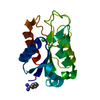
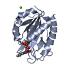
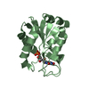
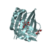
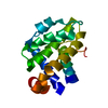
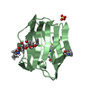
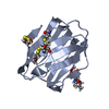
 Helicobacter pylori (bacteria)
 X-RAY DIFFRACTION / Resolution: 2.4 Å
  Authors
 Citation
 Structure visualization
 Downloads & links
 Other downloads
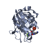

 PDBj
PDBj

 Assembly

 Mass: 456.344 Da / Num. of mol.: 1 / Source method: obtained synthetically / Formula: C17H21N4O9P
 Mass: 18.015 Da / Num. of mol.: 32 / Source method: isolated from a natural source / Formula: H2O
 Sample preparation
 Processing