 Movie
Movie Controller

[English] 日本語
 Yorodumi
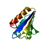
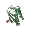
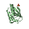
- PDB-1fu0: CRYSTAL STRUCTURE ANALYSIS OF THE PHOSPHO-SERINE 46 HPR FROM ENTE... -
+ Open data

- Basic information
| Entry | Database: PDB / ID: 1fu0 | ||||||
|---|---|---|---|---|---|---|---|
| Title | CRYSTAL STRUCTURE ANALYSIS OF THE PHOSPHO-SERINE 46 HPR FROM ENTEROCOCCUS FAECALIS | ||||||
 Components Components | PHOSPHOCARRIER PROTEIN HPR | ||||||
 Keywords Keywords | SIGNALING PROTEIN / Phospho-Serine HPr / PTS System | ||||||
| Function / homology |  Function and homology information Function and homology informationphosphoenolpyruvate-dependent sugar phosphotransferase system / cytoplasm Similarity search - Function | ||||||
| Biological species |   Enterococcus faecalis (bacteria) Enterococcus faecalis (bacteria) | ||||||
| Method |  X-RAY DIFFRACTION / SYNCHROTRON / Resolution: 1.9 Å X-RAY DIFFRACTION / SYNCHROTRON / Resolution: 1.9 Å | ||||||
 Authors Authors | Audette, G.F. / Engelmann, R. / Hengstenberg, W. / Deutscher, J. / Hayakawa, K. / Quail, J.W. / Delbaere, L.T.J. | ||||||
 Citation Citation | Journal: J.Mol.Biol. / Year: 2000 Title: The 1.9 A resolution structure of phospho-serine 46 HPr from Enterococcus faecalis. Authors: Audette, G.F. / Engelmann, R. / Hengstenberg, W. / Deutscher, J. / Hayakawa, K. / Quail, J.W. / Delbaere, L.T. | ||||||
| History |
|
- Structure visualization
| Structure viewer | Molecule: MolmilJmol/JSmol |
|---|
- Downloads & links
-Download
| PDBx/mmCIF format | 1fu0.cif.gz | 45.2 KB | Display | PDBx/mmCIF format |
|---|---|---|---|---|
| PDB format | pdb1fu0.ent.gz | 31.8 KB | Display | PDB format |
| PDBx/mmJSON format | 1fu0.json.gz | Tree view | PDBx/mmJSON format | |
| Others |  Other downloads Other downloads |
-Validation report
| Arichive directory | https://data.pdbj.org/pub/pdb/validation_reports/fu/1fu0ftp://data.pdbj.org/pub/pdb/validation_reports/fu/1fu0 | HTTPS FTP |
|---|
-Related structure data
| Related structure data | |
|---|---|
| Similar structure data |


-Links
 PDBj
PDBj- Assembly
| Deposited unit | 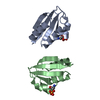
| ||||||||||
|---|---|---|---|---|---|---|---|---|---|---|---|
| 1 | 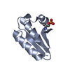
| ||||||||||
| 2 | 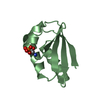
| ||||||||||
| Unit cell |
| ||||||||||
| Noncrystallographic symmetry (NCS) | NCS oper: (Code: given Matrix: (-0.99872, 0.02055, 0.04623), Vector: Details | The biological assembly is a monomer | |
-Components
| #1: Protein | Mass: 9281.466 Da / Num. of mol.: 2 / Source method: isolated from a natural source / Source: (natural) Enterococcus faecalis (bacteria) / Strain: 26487 / References: UniProt: P07515#2: Water | ChemComp-HOH / |  Mass: 18.015 Da / Num. of mol.: 91 / Source method: isolated from a natural source / Formula: H2O Mass: 18.015 Da / Num. of mol.: 91 / Source method: isolated from a natural source / Formula: H2OHas protein modification | Y | |
|---|
-Experimental details
-Experiment
| Experiment | Method: X-RAY DIFFRACTION / Number of used crystals: 1 |
|---|
- Sample preparation
| Crystal | Density Matthews: 1.8 Å3/Da / Density % sol: 31.74 % | ||||||||||||||||||||
|---|---|---|---|---|---|---|---|---|---|---|---|---|---|---|---|---|---|---|---|---|---|
| Crystal grow | Temperature: 287 K / Method: vapor diffusion, hanging drop / pH: 4.2 Details: 2.8M sodium-potassium phosphate, pH 4.2, VAPOR DIFFUSION, HANGING DROP, temperature 287.0K | ||||||||||||||||||||
| Crystal | *PLUS Density % sol: 31.3 % | ||||||||||||||||||||
| Crystal grow | *PLUS Details: used seeding | ||||||||||||||||||||
| Components of the solutions | *PLUS
|
-Data collection
| Diffraction | Mean temperature: 290 K |
|---|---|
| Diffraction source | Source: SYNCHROTRON / Site: Photon Factory  / Beamline: BL-18B / Wavelength: 1 / Beamline: BL-18B / Wavelength: 1 |
| Detector | Type: WEISSENBERG / Detector: DIFFRACTOMETER / Date: Dec 14, 1998 |
| Radiation | Protocol: SINGLE WAVELENGTH / Monochromatic (M) / Laue (L): M / Scattering type: x-ray |
| Radiation wavelength | Wavelength: 1 Å / Relative weight: 1 |
| Reflection | Resolution: 1.9→40 Å / Num. all: 10043 / Num. obs: 10043 / % possible obs: 95.1 % / Redundancy: 4.23 % / Biso Wilson estimate: 14.8 Å2 / Rmerge(I) obs: 0.041 / Net I/σ(I): 8.6 |
| Reflection shell | Resolution: 1.9→1.93 Å / Redundancy: 3.7 % / Rmerge(I) obs: 0.11 / Num. unique all: 447 / % possible all: 84.3 |
| Reflection | *PLUS Num. measured all: 42471 |
| Reflection shell | *PLUS % possible obs: 84.3 % / Num. unique obs: 447 / Num. measured obs: 1668 |
- Processing
| Software |
| |||||||||||||||||||||||||
|---|---|---|---|---|---|---|---|---|---|---|---|---|---|---|---|---|---|---|---|---|---|---|---|---|---|---|
| Refinement | Resolution: 1.9→40 Å / Cross valid method: THROUGHOUT / σ(F): 0 / σ(I): 0 / Stereochemistry target values: Engh & Huber / Details: Simulated Annealing
| |||||||||||||||||||||||||
| Refinement step | Cycle: LAST / Resolution: 1.9→40 Å
| |||||||||||||||||||||||||
| Refine LS restraints |
| |||||||||||||||||||||||||
| Software | *PLUS Name: X-PLOR / Version: 3.851 / Classification: refinement | |||||||||||||||||||||||||
| Refinement | *PLUS Lowest resolution: 40 Å / σ(F): 0 / % reflection Rfree: 5 % | |||||||||||||||||||||||||
| Solvent computation | *PLUS | |||||||||||||||||||||||||
| Displacement parameters | *PLUS | |||||||||||||||||||||||||
| Refine LS restraints | *PLUS
|
