 Movie
Movie Controller

[English] 日本語
 Yorodumi
- PDB-1fr8: CRYSTAL STRUCTURE OF THE BOVINE BETA 1,4 GALACTOSYLTRANSFERASE (B... -
+ Open data

- Basic information
| Entry | Database: PDB / ID: 1fr8 | ||||||
|---|---|---|---|---|---|---|---|
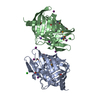
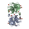
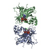
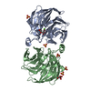
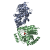
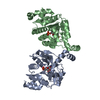
| Title | CRYSTAL STRUCTURE OF THE BOVINE BETA 1,4 GALACTOSYLTRANSFERASE (B4GALT1) CATALYTIC DOMAIN COMPLEXED WITH URIDINE DIPHOSPHOGALACTOSE | ||||||
 Components Components | BETA 1,4 GALACTOSYLTRANSFERASE | ||||||
 Keywords Keywords | TRANSFERASE / nucleotide binding protein alpha beta alpha fold | ||||||
| Function / homology |  Function and homology information Function and homology informationKeratan sulfate biosynthesis / Interaction With Cumulus Cells And The Zona Pellucida / Lactose synthesis / N-Glycan antennae elongation / carbohydrate derivative biosynthetic process / lactose synthase / neolactotriaosylceramide beta-1,4-galactosyltransferase / beta-N-acetylglucosaminylglycopeptide beta-1,4-galactosyltransferase / N-acetyllactosamine synthase / N-acetyllactosamine synthase activity ...Keratan sulfate biosynthesis / Interaction With Cumulus Cells And The Zona Pellucida / Lactose synthesis / N-Glycan antennae elongation / carbohydrate derivative biosynthetic process / lactose synthase / neolactotriaosylceramide beta-1,4-galactosyltransferase / beta-N-acetylglucosaminylglycopeptide beta-1,4-galactosyltransferase / N-acetyllactosamine synthase / N-acetyllactosamine synthase activity / positive regulation of circulating fibrinogen levels / beta-N-acetylglucosaminylglycopeptide beta-1,4-galactosyltransferase activity / penetration of zona pellucida / UDP-galactosyltransferase activity / regulation of acrosome reaction / Golgi trans cisterna / lactose synthase activity / lactose biosynthetic process / macrophage migration / oligosaccharide biosynthetic process / development of animal secondary sexual characteristics / desmosome / acute inflammatory response / galactose metabolic process / positive regulation of epithelial cell proliferation involved in wound healing / binding of sperm to zona pellucida / protein N-linked glycosylation / angiogenesis involved in wound healing / Neutrophil degranulation / Transferases; Glycosyltransferases; Hexosyltransferases / Golgi cisterna membrane / epithelial cell development / alpha-tubulin binding / beta-tubulin binding / extracellular matrix organization / epithelial cell proliferation / lipid metabolic process / filopodium / brush border membrane / negative regulation of epithelial cell proliferation / manganese ion binding / basolateral plasma membrane / cell adhesion / positive regulation of apoptotic process / external side of plasma membrane / Golgi apparatus / protein-containing complex / : / identical protein binding Similarity search - Function | ||||||
| Biological species |  | ||||||
| Method |  X-RAY DIFFRACTION / SYNCHROTRON / Resolution: 2.4 Å X-RAY DIFFRACTION / SYNCHROTRON / Resolution: 2.4 Å | ||||||
 Authors Authors | Gastinel, L.N. / Cambillau, C. / Bourne, Y. | ||||||
 Citation Citation | Journal: EMBO J. / Year: 1999 Title: Crystal structures of the bovine beta4galactosyltransferase catalytic domain and its complex with uridine diphosphogalactose. Authors: Gastinel, L.N. / Cambillau, C. / Bourne, Y. | ||||||
| History |
|
- Structure visualization
| Structure viewer | Molecule: MolmilJmol/JSmol |
|---|
- Downloads & links
-Download
| PDBx/mmCIF format | 1fr8.cif.gz | 122.6 KB | Display | PDBx/mmCIF format |
|---|---|---|---|---|
| PDB format | pdb1fr8.ent.gz | 96.2 KB | Display | PDB format |
| PDBx/mmJSON format | 1fr8.json.gz | Tree view | PDBx/mmJSON format | |
| Others |  Other downloads Other downloads |
-Validation report
| Arichive directory | https://data.pdbj.org/pub/pdb/validation_reports/fr/1fr8ftp://data.pdbj.org/pub/pdb/validation_reports/fr/1fr8 | HTTPS FTP |
|---|
-Related structure data


-Links
 PDBj
PDBj

- Assembly
| Deposited unit | 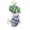
| ||||||||
|---|---|---|---|---|---|---|---|---|---|
| 1 |
| ||||||||
| Unit cell |
|
-Components
| #1: Protein | Mass: 33072.824 Da / Num. of mol.: 2 / Fragment: CATALYTIC DOMAIN Source method: isolated from a genetically manipulated source Source: (gene. exp.) References: UniProt: P08037, beta-N-acetylglucosaminylglycopeptide beta-1,4-galactosyltransferase #2: Chemical |   Mass: 566.302 Da / Num. of mol.: 2 / Source method: obtained synthetically / Formula: C15H24N2O17P2 Mass: 566.302 Da / Num. of mol.: 2 / Source method: obtained synthetically / Formula: C15H24N2O17P2#3: Water | ChemComp-HOH / |  Mass: 18.015 Da / Num. of mol.: 79 / Source method: isolated from a natural source / Formula: H2O Mass: 18.015 Da / Num. of mol.: 79 / Source method: isolated from a natural source / Formula: H2OHas protein modification | Y | |
|---|
-Experimental details
-Experiment
| Experiment | Method: X-RAY DIFFRACTION / Number of used crystals: 1 |
|---|
- Sample preparation
| Crystal | Density Matthews: 3.39 Å3/Da / Density % sol: 63.75 % | ||||||||||||||||||||||||||||||||||||||||||||||||
|---|---|---|---|---|---|---|---|---|---|---|---|---|---|---|---|---|---|---|---|---|---|---|---|---|---|---|---|---|---|---|---|---|---|---|---|---|---|---|---|---|---|---|---|---|---|---|---|---|---|
| Crystal grow | Temperature: 293 K / Method: vapor diffusion, hanging drop / pH: 8.5 Details: PEG 5000, Tris, MnCl2, Li2SO4, pH 8.5, VAPOR DIFFUSION, HANGING DROP, temperature 293K | ||||||||||||||||||||||||||||||||||||||||||||||||
| Crystal grow | *PLUS Temperature: 20 ℃ / pH: 6.5 | ||||||||||||||||||||||||||||||||||||||||||||||||
| Components of the solutions | *PLUS
|
-Data collection
| Diffraction | Mean temperature: 100 K |
|---|---|
| Diffraction source | Source: SYNCHROTRON / Site: ESRF  / Beamline: ID14-1 / Wavelength: 0.933 / Beamline: ID14-1 / Wavelength: 0.933 |
| Detector | Type: CUSTOM-MADE / Detector: CCD / Date: Sep 17, 1998 |
| Radiation | Protocol: SINGLE WAVELENGTH / Monochromatic (M) / Laue (L): M / Scattering type: x-ray |
| Radiation wavelength | Wavelength: 0.933 Å / Relative weight: 1 |
| Reflection | Resolution: 2.4→30 Å / Num. obs: 32664 / % possible obs: 91.9 % / Observed criterion σ(F): 0 / Observed criterion σ(I): 0 / Redundancy: 4.3 % / Biso Wilson estimate: 49.8 Å2 / Rmerge(I) obs: 0.065 / Net I/σ(I): 7.2 |
| Reflection shell | Resolution: 2.4→2.5 Å / Redundancy: 4.1 % / Rmerge(I) obs: 0.425 / % possible all: 92.6 |
| Reflection | *PLUS Num. measured all: 138168 |
| Reflection shell | *PLUS % possible obs: 94 % / Mean I/σ(I) obs: 2.1 |
- Processing
| Software |
| |||||||||||||||||||||||||
|---|---|---|---|---|---|---|---|---|---|---|---|---|---|---|---|---|---|---|---|---|---|---|---|---|---|---|
| Refinement | Resolution: 2.4→30 Å / Cross valid method: FREE R-VALUE / σ(F): 0 / σ(I): 0 / Stereochemistry target values: CNS / Details: maximum likelihood target using amplitudes
| |||||||||||||||||||||||||
| Refinement step | Cycle: LAST / Resolution: 2.4→30 Å
| |||||||||||||||||||||||||
| Refine LS restraints |
| |||||||||||||||||||||||||
| Software | *PLUS Name: CNS / Version: 0.9 / Classification: refinement | |||||||||||||||||||||||||
| Refinement | *PLUS Highest resolution: 2.4 Å / Lowest resolution: 30 Å / σ(F): 0 / % reflection Rfree: 3 % | |||||||||||||||||||||||||
| Solvent computation | *PLUS | |||||||||||||||||||||||||
| Displacement parameters | *PLUS | |||||||||||||||||||||||||
| Refine LS restraints | *PLUS Type: c_angle_deg / Dev ideal: 2.1 |
