 Movie
Movie Controller

[English] 日本語
 Yorodumi
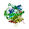
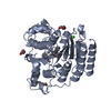
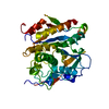
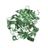
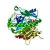
- PDB-1fml: CRYSTAL STRUCTURE OF RETINOL DEHYDRATASE IN A COMPLEX WITH RETINO... -
+ Open data

- Basic information
| Entry | Database: PDB / ID: 1fml | ||||||
|---|---|---|---|---|---|---|---|
| Title | CRYSTAL STRUCTURE OF RETINOL DEHYDRATASE IN A COMPLEX WITH RETINOL AND PAP | ||||||
 Components Components | RETINOL DEHYDRATASE | ||||||
 Keywords Keywords | TRANSFERASE / sulfotransferase / dehydratase / retinol / adenosine 3' / 5'-diphosphate | ||||||
| Function / homology |  Function and homology information Function and homology information | ||||||
| Biological species |   Spodoptera frugiperda (fall armyworm) Spodoptera frugiperda (fall armyworm) | ||||||
| Method |  X-RAY DIFFRACTION / SIRAS Phasing / Resolution: 2.75 Å X-RAY DIFFRACTION / SIRAS Phasing / Resolution: 2.75 Å | ||||||
 Authors Authors | Pakhomova, S. / Kobayashi, M. / Buck, J. / Newcomer, M.E. | ||||||
 Citation Citation | Journal: Nat.Struct.Biol. / Year: 2001 Title: A helical lid converts a sulfotransferase to a dehydratase. Authors: Pakhomova, S. / Kobayashi, M. / Buck, J. / Newcomer, M.E. | ||||||
| History |
|
- Structure visualization
| Structure viewer | Molecule: MolmilJmol/JSmol |
|---|
- Downloads & links
-Download
| PDBx/mmCIF format | 1fml.cif.gz | 141.5 KB | Display | PDBx/mmCIF format |
|---|---|---|---|---|
| PDB format | pdb1fml.ent.gz | 113 KB | Display | PDB format |
| PDBx/mmJSON format | 1fml.json.gz | Tree view | PDBx/mmJSON format | |
| Others |  Other downloads Other downloads |
-Validation report
| Arichive directory | https://data.pdbj.org/pub/pdb/validation_reports/fm/1fmlftp://data.pdbj.org/pub/pdb/validation_reports/fm/1fml | HTTPS FTP |
|---|
-Related structure data


-Links
 PDBj
PDBj
- Assembly
| Deposited unit | 
| ||||||||
|---|---|---|---|---|---|---|---|---|---|
| 1 | 
| ||||||||
| 2 | 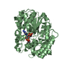
| ||||||||
| Unit cell |
|
-Components
| #1: Protein | Mass: 41555.480 Da / Num. of mol.: 2 Source method: isolated from a genetically manipulated source Source: (gene. exp.) Spodoptera frugiperda (fall armyworm) / Plasmid: PET-15B / Production host:  #2: Chemical |   Mass: 40.078 Da / Num. of mol.: 2 / Source method: obtained synthetically / Formula: Ca Mass: 40.078 Da / Num. of mol.: 2 / Source method: obtained synthetically / Formula: Ca#3: Chemical | 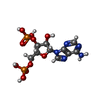  Type: RNA linking / Mass: 427.201 Da / Num. of mol.: 2 / Source method: obtained synthetically / Formula: C10H15N5O10P2 Type: RNA linking / Mass: 427.201 Da / Num. of mol.: 2 / Source method: obtained synthetically / Formula: C10H15N5O10P2#4: Chemical |   Mass: 286.452 Da / Num. of mol.: 2 / Source method: obtained synthetically / Formula: C20H30O Mass: 286.452 Da / Num. of mol.: 2 / Source method: obtained synthetically / Formula: C20H30O#5: Water | ChemComp-HOH / |  Mass: 18.015 Da / Num. of mol.: 91 / Source method: isolated from a natural source / Formula: H2O Mass: 18.015 Da / Num. of mol.: 91 / Source method: isolated from a natural source / Formula: H2O |
|---|
-Experimental details
-Experiment
| Experiment | Method: X-RAY DIFFRACTION / Number of used crystals: 1 |
|---|
- Sample preparation
| Crystal | Density Matthews: 2.6 Å3/Da / Density % sol: 51.6 % | ||||||||||||||||||||||||||||||
|---|---|---|---|---|---|---|---|---|---|---|---|---|---|---|---|---|---|---|---|---|---|---|---|---|---|---|---|---|---|---|---|
| Crystal grow | Temperature: 284 K / Method: vapor diffusion, hanging drop / pH: 6.6 Details: PEG 3350, calcium chloride, Hepes, glycerol, pH 6.6, VAPOR DIFFUSION, HANGING DROP, temperature 284K | ||||||||||||||||||||||||||||||
| Crystal grow | *PLUS Details: Pakhomova, S., (2000) Acta Crystallogr.D56, 1641. / PH range low: 6.8 / PH range high: 6.6 | ||||||||||||||||||||||||||||||
| Components of the solutions | *PLUS
|
-Data collection
| Diffraction | Mean temperature: 100 K |
|---|---|
| Diffraction source | Source: ROTATING ANODE / Type: RIGAKU RU200 / Wavelength: 1.5418 |
| Detector | Type: RIGAKU RAXIS IV / Detector: IMAGE PLATE / Date: Apr 14, 1999 |
| Radiation | Protocol: SINGLE WAVELENGTH / Monochromatic (M) / Laue (L): M / Scattering type: x-ray |
| Radiation wavelength | Wavelength: 1.5418 Å / Relative weight: 1 |
| Reflection | Resolution: 2.75→40 Å / Num. all: 64864 / Num. obs: 20388 / % possible obs: 92.6 % / Observed criterion σ(F): 0 / Observed criterion σ(I): -3 / Redundancy: 3.2 % / Biso Wilson estimate: 21.9 Å2 / Rsym value: 0.104 / Net I/σ(I): 12.7 |
| Reflection shell | Resolution: 2.75→2.85 Å / Redundancy: 2.8 % / Mean I/σ(I) obs: 3.2 / Num. unique all: 2136 / Rsym value: 0.371 / % possible all: 98.9 |
| Reflection | *PLUS Rmerge(I) obs: 0.104 |
| Reflection shell | *PLUS % possible obs: 98.9 % / Rmerge(I) obs: 0.371 |
- Processing
| Software |
| |||||||||||||||||||||||||
|---|---|---|---|---|---|---|---|---|---|---|---|---|---|---|---|---|---|---|---|---|---|---|---|---|---|---|
| Refinement | Method to determine structure: SIRAS Phasing / Resolution: 2.75→38.45 Å / Rfactor Rfree error: 0.006 / Data cutoff high rms absF: 151607.03 / Isotropic thermal model: GROUP / Cross valid method: THROUGHOUT / σ(F): 0 / σ(I): 0 / Stereochemistry target values: Engh & Huber
| |||||||||||||||||||||||||
| Solvent computation | Solvent model: flat model / Bsol: 28.24 Å2 / ksol: 0.315288 e/Å3 | |||||||||||||||||||||||||
| Displacement parameters | Biso mean: 33.6 Å2
| |||||||||||||||||||||||||
| Refine analyze |
| |||||||||||||||||||||||||
| Refinement step | Cycle: LAST / Resolution: 2.75→38.45 Å
| |||||||||||||||||||||||||
| Refine LS restraints |
| |||||||||||||||||||||||||
| LS refinement shell | Resolution: 2.75→2.85 Å / Rfactor Rfree error: 0.035 / Total num. of bins used: 10
| |||||||||||||||||||||||||
| Xplor file |
| |||||||||||||||||||||||||
| Software | *PLUS Name: CNS / Version: 0.9 / Classification: refinement | |||||||||||||||||||||||||
| Refine LS restraints | *PLUS
|
