 Movie
Movie Controller


+ Open data

- Basic information
| Entry | Database: PDB / ID: 1flh | ||||||
|---|---|---|---|---|---|---|---|
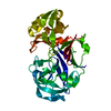
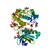
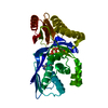
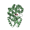
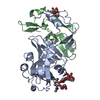
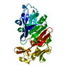
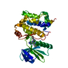
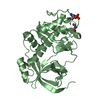
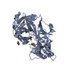
| Title | CRYSTAL STRUCTURE OF HUMAN UROPEPSIN AT 2.45 A RESOLUTION | ||||||
 Components Components | UROPEPSIN | ||||||
 Keywords Keywords | HYDROLASE / ACID PROTEINASE | ||||||
| Function / homology |  Function and homology information Function and homology informationmultivesicular body lumen / pepsin A / Surfactant metabolism / digestion / aspartic-type endopeptidase activity / proteolysis / extracellular exosome Similarity search - Function | ||||||
| Biological species |  Homo sapiens (human) Homo sapiens (human) | ||||||
| Method |  X-RAY DIFFRACTION / SYNCHROTRON / MOLECULAR REPLACEMENT / Resolution: 2.45 Å X-RAY DIFFRACTION / SYNCHROTRON / MOLECULAR REPLACEMENT / Resolution: 2.45 Å | ||||||
 Authors Authors | Canduri, F. / Teodoro, L.G.V.L. / Fadel, V. / Lorenzi, C.C.B. / Hial, V. / Gomes, R.A.S. / Neto, J.R. / De Azevedo Jr., W.F. | ||||||
 Citation Citation | Journal: Acta Crystallogr.,Sect.D / Year: 2001 Title: Structure of human uropepsin at 2.45 A resolution. Authors: Canduri, F. / Teodoro, L.G. / Fadel, V. / Lorenzi, C.C. / Hial, V. / Gomes, R.A. / Neto, J.R. / de Azevedo, W.F. #1: Journal: BIOCHEM.MOL.BIOL.INT. / Year: 1998Title: Crystallization, Preliminary X-Ray Analysis and Patterson Search of a New Aspartic Protease Isolate from Human Urine Authors: Canduri, F. / Teodoro, L.G.V.L. / Lorenzi, C.C.B. / Gomes, R.A.S. / Fontes, M.R.M. / Arni, R.K. / De Azevedo Jr., W.F. | ||||||
| History |
|
- Structure visualization
| Structure viewer | Molecule: MolmilJmol/JSmol |
|---|
- Downloads & links
-Download
| PDBx/mmCIF format | 1flh.cif.gz | 74.5 KB | Display | PDBx/mmCIF format |
|---|---|---|---|---|
| PDB format | pdb1flh.ent.gz | 54.9 KB | Display | PDB format |
| PDBx/mmJSON format | 1flh.json.gz | Tree view | PDBx/mmJSON format | |
| Others |  Other downloads Other downloads |
-Validation report
| Arichive directory | https://data.pdbj.org/pub/pdb/validation_reports/fl/1flhftp://data.pdbj.org/pub/pdb/validation_reports/fl/1flh | HTTPS FTP |
|---|
-Related structure data
| Related structure data |  1psnS S: Starting model for refinement |
|---|---|
| Similar structure data |

-Links
 PDBj
PDBj
- Assembly
| Deposited unit | 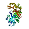
| ||||||||
|---|---|---|---|---|---|---|---|---|---|
| 1 |
| ||||||||
| Unit cell |
|
-Components
| #1: Protein | Mass: 34617.859 Da / Num. of mol.: 1 / Source method: isolated from a natural source / Source: (natural) Homo sapiens (human) / References: UniProt: P00790, UniProt: P0DJD7*PLUS, pepsin A |
|---|---|
| #2: Water | ChemComp-HOH /  Mass: 18.015 Da / Num. of mol.: 143 / Source method: isolated from a natural source / Formula: H2O Mass: 18.015 Da / Num. of mol.: 143 / Source method: isolated from a natural source / Formula: H2O |
| Has protein modification | Y |
-Experimental details
-Experiment
| Experiment | Method: X-RAY DIFFRACTION / Number of used crystals: 1 |
|---|
- Sample preparation
| Crystal | Density Matthews: 2.17 Å3/Da / Density % sol: 43.3 % | ||||||||||||||||||||
|---|---|---|---|---|---|---|---|---|---|---|---|---|---|---|---|---|---|---|---|---|---|
| Crystal grow | Method: vapor diffusion / pH: 7 Details: 0.1M HEPES BUFFER (pH 7.0), 2% PEG 400, 2.0M AMMONIUM SULFATE., VAPOR DIFFUSION | ||||||||||||||||||||
| Crystal grow | *PLUS Method: vapor diffusion, hanging drop | ||||||||||||||||||||
| Components of the solutions | *PLUS
|
-Data collection
| Diffraction source | Source: SYNCHROTRON / Site: LNLS  / Beamline: D03B-MX1 / Wavelength: 1.38 / Beamline: D03B-MX1 / Wavelength: 1.38 |
|---|---|
| Detector | Type: MARRESEARCH / Detector: IMAGE PLATE / Date: Jul 12, 1998 |
| Radiation | Protocol: SINGLE WAVELENGTH / Monochromatic (M) / Laue (L): M / Scattering type: x-ray |
| Radiation wavelength | Wavelength: 1.38 Å / Relative weight: 1 |
| Reflection | Resolution: 2.45→12 Å / Num. obs: 13134 / % possible obs: 98.9 % / Observed criterion σ(I): 1 / Redundancy: 3.26 % / Rmerge(I) obs: 0.077 |
| Reflection | *PLUS Num. measured all: 24189 |
| Reflection shell | *PLUS Highest resolution: 2.45 Å / Lowest resolution: 2.51 Å / % possible obs: 99.1 % / Rmerge(I) obs: 0.303 |
- Processing
| Software |
| |||||||||||||||
|---|---|---|---|---|---|---|---|---|---|---|---|---|---|---|---|---|
| Refinement | Method to determine structure: MOLECULAR REPLACEMENT Starting model: 1PSN Resolution: 2.45→12 Å / σ(F): 2
| |||||||||||||||
| Displacement parameters | Biso mean: 15.52 Å2 | |||||||||||||||
| Refine analyze | Luzzati coordinate error obs: 0.19 Å | |||||||||||||||
| Refinement step | Cycle: LAST / Resolution: 2.45→12 Å
| |||||||||||||||
| Refine LS restraints |
| |||||||||||||||
| Software | *PLUS Name: X-PLOR / Classification: refinement | |||||||||||||||
| Refinement | *PLUS Lowest resolution: 12 Å / σ(F): 2 / % reflection Rfree: 10 % / Rfactor Rfree: 0.251 | |||||||||||||||
| Solvent computation | *PLUS | |||||||||||||||
| Displacement parameters | *PLUS | |||||||||||||||
| Refine LS restraints | *PLUS
|
