[English] 日本語
Yorodumi
- PDB-1fl2: CATALYTIC CORE COMPONENT OF THE ALKYLHYDROPEROXIDE REDUCTASE AHPF... -

+
Open data


ID or keywords:

Loading...

-
Basic information

Entry
Database: PDB / ID: 1fl2
TitleCATALYTIC CORE COMPONENT OF THE ALKYLHYDROPEROXIDE REDUCTASE AHPF FROM E.COLI
ComponentsALKYL HYDROPEROXIDE REDUCTASE SUBUNIT F
KeywordsOXIDOREDUCTASE / alkylhydroperoxide reductase / reactive oxygen / FAD / disulphide oxidoreductase
Function / homology
Function and homology information


alkyl hydroperoxide reductase complex / Oxidoreductases; Acting on a sulfur group of donors; With NAD+ or NADP+ as acceptor / alkyl hydroperoxide reductase activity / thioredoxin-disulfide reductase (NADPH) activity / FAD binding / response to reactive oxygen species / cell redox homeostasis / hydrogen peroxide catabolic process / NAD binding / response to oxidative stress / cytosol
Similarity search - Function
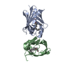
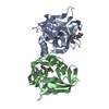
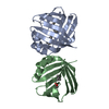
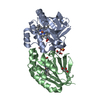
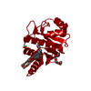
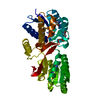
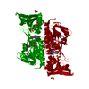
Alkyl hydroperoxide reductase subunit F / AhpF, N-terminal domain, C-terminal TRX-fold subdomain / AhpF, N-terminal domain, N-terminal TRX-fold subdomain / Thioredoxin domain / Pyridine nucleotide-disulphide oxidoreductase, class-II, active site / Pyridine nucleotide-disulphide oxidoreductases class-II active site. / Pyridine nucleotide-disulphide oxidoreductase / Thioredoxin-like fold / Glutaredoxin domain profile. / FAD/NAD(P)-binding domain ...Alkyl hydroperoxide reductase subunit F / AhpF, N-terminal domain, C-terminal TRX-fold subdomain / AhpF, N-terminal domain, N-terminal TRX-fold subdomain / Thioredoxin domain / Pyridine nucleotide-disulphide oxidoreductase, class-II, active site / Pyridine nucleotide-disulphide oxidoreductases class-II active site. / Pyridine nucleotide-disulphide oxidoreductase / Thioredoxin-like fold / Glutaredoxin domain profile. / FAD/NAD(P)-binding domain / Pyridine nucleotide-disulphide oxidoreductase / FAD/NAD(P)-binding domain / FAD/NAD(P)-binding domain / 3-Layer(bba) Sandwich / FAD/NAD(P)-binding domain superfamily / Thioredoxin-like superfamily / Alpha Beta
Similarity search - Domain/homology
FLAVIN-ADENINE DINUCLEOTIDE / Alkyl hydroperoxide reductase subunit F
Similarity search - Component
Biological speciesEscherichia coli (E. coli)
MethodX-RAY DIFFRACTION / SYNCHROTRON / Resolution: 1.9 Å
AuthorsBieger, B. / Essen, L.-O.
CitationJournal: J.Mol.Biol. / Year: 2001
Title: Crystal structure of the catalytic core component of the alkylhydroperoxide reductase AhpF from Escherichia coli.
Authors: Bieger, B. / Essen, L.O.
History
DepositionAug 11, 2000Deposition site: RCSB / Processing site: RCSB
Revision 1.0Mar 21, 2001Provider: repository / Type: Initial release
Revision 1.1Apr 27, 2008Group: Version format compliance
Revision 1.2Jul 13, 2011Group: Derived calculations / Version format compliance
Revision 1.3Jan 31, 2018Group: Experimental preparation / Category: exptl_crystal_grow / Item: _exptl_crystal_grow.temp

-
Structure visualization

Structure viewerMolecule:
MolmilJmol/JSmol

Downloads & links

-
Assembly

Deposited unit
A: ALKYL HYDROPEROXIDE REDUCTASE SUBUNIT F
hetero molecules


Theoretical massNumber of molelcules
Total (without water)34,1977
Polymers32,9321
Non-polymers1,2666
Water7,368409
1


  • Idetical with deposited unit
  • defined by author&software
TypeNameSymmetry operationNumber
identity operation1_555x,y,z1
MethodPQS
2
A: ALKYL HYDROPEROXIDE REDUCTASE SUBUNIT F
hetero molecules

A: ALKYL HYDROPEROXIDE REDUCTASE SUBUNIT F
hetero molecules


Theoretical massNumber of molelcules
Total (without water)68,39514
Polymers65,8632
Non-polymers2,53212
Water362
TypeNameSymmetry operationNumber
identity operation1_555x,y,z1
crystal symmetry operation4_555y,x,-z1
Buried area9000 Å2
ΔGint-148 kcal/mol
Surface area24340 Å2
MethodPISA
Unit cell
Length a, b, c (Å)60.440, 60.440, 171.830
Angle α, β, γ (deg.)90.00, 90.00, 120.00
Int Tables number154
Space group name H-MP3221
Components on special symmetry positions
IDModelComponents
11A-840-

HOH

21A-877-

HOH

-
Components

#1: Protein ALKYL HYDROPEROXIDE REDUCTASE SUBUNIT F / ALKYL HYDROPEROXIDE REDUCTASE F52A PROTEIN


Mass: 32931.531 Da / Num. of mol.: 1 / Fragment: C-TERMINAL DOMAIN
Source method: isolated from a genetically manipulated source
Source: (gene. exp.) Escherichia coli (E. coli) / Production host: Escherichia coli (E. coli)
References: UniProt: P35340, Oxidoreductases; Acting on a sulfur group of donors; With NAD+ or NADP+ as acceptor
#2: Chemical
ChemComp-SO4 / SULFATE ION / Sulfate


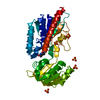
Mass: 96.063 Da / Num. of mol.: 5 / Source method: obtained synthetically / Formula: SO4
#3: Chemical ChemComp-FAD / FLAVIN-ADENINE DINUCLEOTIDE / Flavin adenine dinucleotide


Mass: 785.550 Da / Num. of mol.: 1 / Source method: obtained synthetically / Formula: C27H33N9O15P2 / Comment: FAD*YM
#4: Water ChemComp-HOH / water / Water


Mass: 18.015 Da / Num. of mol.: 409 / Source method: isolated from a natural source / Formula: H2O

-
Experimental details

-
Experiment

ExperimentMethod: X-RAY DIFFRACTION / Number of used crystals: 1

-
Sample preparation

CrystalDensity Matthews: 2.75 Å3/Da / Density % sol: 55.28 %
Crystal growTemperature: 291 K / Method: vapor diffusion, hanging drop / pH: 6.5
Details: 2.0 M ammonium sulfate, 6% PEG 400, 0.1 M Tris/acetate , pH 6.5, VAPOR DIFFUSION, HANGING DROP, temperature 18K
Crystal grow
*PLUS
Temperature: 291 K / pH: 8 / Details: Bieger, B., (2000) Acta Crystallogr. D56, 92.
Components of the solutions
*PLUS
IDConc.Common nameCrystal-IDSol-ID
17 mg/mlprotein1drop
25 mMTris-HCl1drop
32.0 Mammonium sulfate1reservoir
46 %PEG4001reservoir
50.1 MTris-acetate1reservoir

-
Data collection

DiffractionMean temperature: 100 K
Diffraction sourceSource: SYNCHROTRON / Site: MPG/DESY, HAMBURG / Beamline: BW6 / Wavelength: 1
DetectorType: MARRESEARCH / Detector: CCD / Date: Dec 6, 1998
RadiationProtocol: SINGLE WAVELENGTH / Monochromatic (M) / Laue (L): M / Scattering type: x-ray
Radiation wavelengthWavelength: 1 Å / Relative weight: 1
ReflectionResolution: 1.9→25 Å / Num. all: 29657 / Num. obs: 126257 / % possible obs: 99.6 % / Observed criterion σ(F): 0 / Observed criterion σ(I): 0 / Redundancy: 4 % / Biso Wilson estimate: 10.9 Å2 / Rmerge(I) obs: 0.045 / Net I/σ(I): 20.9
Reflection shellResolution: 1.9→1.95 Å / Redundancy: 3 % / Rmerge(I) obs: 0.109 / % possible all: 99.2
Reflection
*PLUS
Num. obs: 29657 / Num. measured all: 126257
Reflection shell
*PLUS
% possible obs: 99.2 % / Mean I/σ(I) obs: 12.3

-
Processing

Software
NameVersionClassification
DENZOdata reduction
SCALEPACKdata scaling
AMoREphasing
CNS0.9refinement
RefinementResolution: 1.9→20 Å / Rfactor Rfree error: 0.006 / Data cutoff high absF: 2267834.21 / Data cutoff low absF: 0 / Isotropic thermal model: RESTRAINED / Cross valid method: THROUGHOUT / σ(F): 0 / σ(I): 0 / Stereochemistry target values: Engh & Huber
RfactorNum. reflection% reflectionSelection details
Rfree0.217 1447 4.9 %RANDOM
Rwork0.197 ---
all0.197 126257 --
obs0.197 29484 99.6 %-
Solvent computationSolvent model: FLAT MODEL / Bsol: 50.86 Å2 / ksol: 0.418 e/Å3
Displacement parametersBiso mean: 18.2 Å2
Baniso -1Baniso -2Baniso -3
1-0.21 Å2-0.31 Å20 Å2
2--0.21 Å20 Å2
3----0.41 Å2
Refine analyze
FreeObs
Luzzati coordinate error0.22 Å0.2 Å
Luzzati d res low-5 Å
Luzzati sigma a0.12 Å0.08 Å
Refinement stepCycle: LAST / Resolution: 1.9→20 Å
ProteinNucleic acidLigandSolventTotal
Num. atoms2311 0 78 409 2798
Refine LS restraints
Refine-IDTypeDev ideal
X-RAY DIFFRACTIONc_bond_d0.011
X-RAY DIFFRACTIONc_angle_deg1.6
X-RAY DIFFRACTIONc_dihedral_angle_d24.9
X-RAY DIFFRACTIONc_improper_angle_d1.53
LS refinement shellResolution: 1.9→2.02 Å / Rfactor Rfree error: 0.015 / Total num. of bins used: 6
RfactorNum. reflection% reflection
Rfree0.225 232 4.8 %
Rwork0.199 4573 -
obs--99.2 %
Xplor file
Refine-IDSerial noParam fileTopol file
X-RAY DIFFRACTION1PROTEIN_REP.PARAMPROTEIN.TOP
X-RAY DIFFRACTION2FAD.PARAMFAD.TOP
X-RAY DIFFRACTION3PARAM.PO4TOP.PO4
X-RAY DIFFRACTION4WATER.PARAMWATER.TOP
Software
*PLUS
Name: CNS / Version: 0.9 / Classification: refinement
Refine LS restraints
*PLUS
Refine-IDTypeDev ideal
X-RAY DIFFRACTIONc_dihedral_angle_d
X-RAY DIFFRACTIONc_dihedral_angle_deg24.9
X-RAY DIFFRACTIONc_improper_angle_d
X-RAY DIFFRACTIONc_improper_angle_deg1.53

+
About Yorodumi

-
News

-
Feb 9, 2022. New format data for meta-information of EMDB entries

New format data for meta-information of EMDB entries

  • Version 3 of the EMDB header file is now the official format.
  • The previous official version 1.9 will be removed from the archive.

Related info.:EMDB header

External links:wwPDB to switch to version 3 of the EMDB data model

-
Aug 12, 2020. Covid-19 info

Covid-19 info

URL: https://pdbj.org/emnavi/covid19.php

New page: Covid-19 featured information page in EM Navigator.

Related info.:Covid-19 info / Mar 5, 2020. Novel coronavirus structure data

+
Mar 5, 2020. Novel coronavirus structure data

Novel coronavirus structure data

Related info.:Yorodumi Speices / Aug 12, 2020. Covid-19 info

External links:COVID-19 featured content - PDBj / Molecule of the Month (242):Coronavirus Proteases

+
Jan 31, 2019. EMDB accession codes are about to change! (news from PDBe EMDB page)

EMDB accession codes are about to change! (news from PDBe EMDB page)

  • The allocation of 4 digits for EMDB accession codes will soon come to an end. Whilst these codes will remain in use, new EMDB accession codes will include an additional digit and will expand incrementally as the available range of codes is exhausted. The current 4-digit format prefixed with “EMD-” (i.e. EMD-XXXX) will advance to a 5-digit format (i.e. EMD-XXXXX), and so on. It is currently estimated that the 4-digit codes will be depleted around Spring 2019, at which point the 5-digit format will come into force.
  • The EM Navigator/Yorodumi systems omit the EMD- prefix.

Related info.:Q: What is EMD? / ID/Accession-code notation in Yorodumi/EM Navigator

External links:EMDB Accession Codes are Changing Soon! / Contact to PDBj

+
Jul 12, 2017. Major update of PDB

Major update of PDB

  • wwPDB released updated PDB data conforming to the new PDBx/mmCIF dictionary.
  • This is a major update changing the version number from 4 to 5, and with Remediation, in which all the entries are updated.
  • In this update, many items about electron microscopy experimental information are reorganized (e.g. em_software).
  • Now, EM Navigator and Yorodumi are based on the updated data.

External links:wwPDB Remediation / Enriched Model Files Conforming to OneDep Data Standards Now Available in the PDB FTP Archive

-
Yorodumi

Thousand views of thousand structures

  • Yorodumi is a browser for structure data from EMDB, PDB, SASBDB, etc.
  • This page is also the successor to EM Navigator detail page, and also detail information page/front-end page for Omokage search.
  • The word "yorodu" (or yorozu) is an old Japanese word meaning "ten thousand". "mi" (miru) is to see.

Related info.:EMDB / PDB / SASBDB / Comparison of 3 databanks / Yorodumi Search / Aug 31, 2016. New EM Navigator & Yorodumi / Yorodumi Papers / Jmol/JSmol / Function and homology information / Changes in new EM Navigator and Yorodumi

Read more