 Movie
Movie Controller

[English] 日本語
 Yorodumi
- PDB-5k90: Crystal structure of dimeric chlorite dismutase from Cyanothece s... -
+ Open data

- Basic information
| Entry | Database: PDB / ID: 5k90 | ||||||
|---|---|---|---|---|---|---|---|
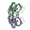
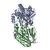
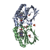
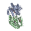
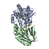
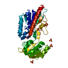
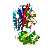
| Title | Crystal structure of dimeric chlorite dismutase from Cyanothece sp. PCC7425 in complex with isothiocyanate | ||||||
 Components Components | Chlorite dismutase | ||||||
 Keywords Keywords | OXIDOREDUCTASE / chlorite dismutase / cyanobacteria / heme / ferredoxin-like fold | ||||||
| Function / homology |  Function and homology information Function and homology informationhydrogen peroxide-dependent heme synthase / oxidoreductase activity / heme binding / metal ion binding Similarity search - Function | ||||||
| Biological species | Cyanothece sp. | ||||||
| Method |  X-RAY DIFFRACTION / SYNCHROTRON / MOLECULAR REPLACEMENT / Resolution: 1.28 Å X-RAY DIFFRACTION / SYNCHROTRON / MOLECULAR REPLACEMENT / Resolution: 1.28 Å | ||||||
 Authors Authors | Puehringer, D. / Schaffner, I. / Mlynek, G. / Obinger, C. / Djinovic-Carugo, K. | ||||||
 Citation Citation | Journal: ACS Catal / Year: 2017 Title: Molecular Mechanism of Enzymatic Chlorite Detoxification: Insights from Structural and Kinetic Studies. Authors: Schaffner, I. / Mlynek, G. / Flego, N. / Puhringer, D. / Libiseller-Egger, J. / Coates, L. / Hofbauer, S. / Bellei, M. / Furtmuller, P.G. / Battistuzzi, G. / Smulevich, G. / Djinovic-Carugo, K. / Obinger, C. | ||||||
| History |
|
- Structure visualization
| Structure viewer | Molecule: MolmilJmol/JSmol |
|---|
- Downloads & links
-Download
| PDBx/mmCIF format | 5k90.cif.gz | 471.6 KB | Display | PDBx/mmCIF format |
|---|---|---|---|---|
| PDB format | pdb5k90.ent.gz | 397.5 KB | Display | PDB format |
| PDBx/mmJSON format | 5k90.json.gz | Tree view | PDBx/mmJSON format | |
| Others |  Other downloads Other downloads |
-Validation report
| Arichive directory | https://data.pdbj.org/pub/pdb/validation_reports/k9/5k90ftp://data.pdbj.org/pub/pdb/validation_reports/k9/5k90 | HTTPS FTP |
|---|
-Related structure data


-Links
 PDBj
PDBj- Assembly
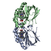
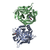
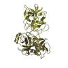
| Deposited unit | 
| ||||||||
|---|---|---|---|---|---|---|---|---|---|
| 1 | 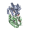
| ||||||||
| 2 | 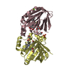
| ||||||||
| Unit cell |
|
-Components
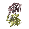
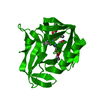
-Protein , 1 types, 4 molecules ABCD
| #1: Protein | Mass: 21830.881 Da / Num. of mol.: 4 Source method: isolated from a genetically manipulated source Source: (gene. exp.)  Cyanothece sp. (strain PCC 7425 / ATCC 29141) (bacteria) Cyanothece sp. (strain PCC 7425 / ATCC 29141) (bacteria)Strain: PCC 7425 / ATCC 29141 / Gene: Cyan7425_1434 / Details (production host): pET-52b+ / Production host: |
|---|
-Non-polymers , 6 types, 821 molecules 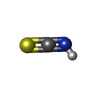


| #2: Chemical | ChemComp-IS8 /  Mass: 59.090 Da / Num. of mol.: 4 / Source method: obtained synthetically / Formula: CHNS Mass: 59.090 Da / Num. of mol.: 4 / Source method: obtained synthetically / Formula: CHNS#3: Chemical | ChemComp-HEM /  Mass: 616.487 Da / Num. of mol.: 4 Mass: 616.487 Da / Num. of mol.: 4Source method: isolated from a genetically manipulated source Formula: C34H32FeN4O4 #4: Chemical | ChemComp-GOL /  Mass: 92.094 Da / Num. of mol.: 9 / Source method: obtained synthetically / Formula: C3H8O3 Mass: 92.094 Da / Num. of mol.: 9 / Source method: obtained synthetically / Formula: C3H8O3#5: Chemical |  Mass: 96.063 Da / Num. of mol.: 2 / Source method: obtained synthetically / Formula: SO4 Mass: 96.063 Da / Num. of mol.: 2 / Source method: obtained synthetically / Formula: SO4#6: Chemical | ChemComp-MG / |  Mass: 24.305 Da / Num. of mol.: 1 / Source method: obtained synthetically / Formula: Mg Mass: 24.305 Da / Num. of mol.: 1 / Source method: obtained synthetically / Formula: Mg#7: Water | ChemComp-HOH / | Mass: 18.015 Da / Num. of mol.: 801 / Source method: isolated from a natural source / Formula: H2O |
|---|
-Experimental details
-Experiment
| Experiment | Method: X-RAY DIFFRACTION / Number of used crystals: 1 |
|---|
- Sample preparation
| Crystal | Density Matthews: 3.04 Å3/Da / Density % sol: 59.6 % |
|---|---|
| Crystal grow | Temperature: 295 K / Method: vapor diffusion, hanging drop / pH: 6.5 Details: 0.15 M MgSO4, 0.1 M MES pH 6.5, 25% (w/v) PEG3350, 10 mM NaSCN |
-Data collection
| Diffraction | Mean temperature: 100 K |
|---|---|
| Diffraction source | Source: SYNCHROTRON / Site: Diamond  / Beamline: I02 / Wavelength: 0.7749 Å / Beamline: I02 / Wavelength: 0.7749 Å |
| Detector | Type: DECTRIS PILATUS 6M / Detector: PIXEL / Date: Dec 3, 2015 Details: Kirkpatrick Baez bimorph mirror pair for horizontal and vertical focussing |
| Radiation | Monochromator: Double crystal Si(111) / Protocol: SINGLE WAVELENGTH / Monochromatic (M) / Laue (L): M / Scattering type: x-ray |
| Radiation wavelength | Wavelength: 0.7749 Å / Relative weight: 1 |
| Reflection | Resolution: 1.28→46.775 Å / Num. obs: 242310 / % possible obs: 93 % / Redundancy: 1.8 % / Biso Wilson estimate: 14.08 Å2 / CC1/2: 0.997 / Rmerge(I) obs: 0.07663 / Net I/σ(I): 4.99 |
| Reflection shell | Resolution: 1.28→1.326 Å / Redundancy: 1.8 % / Rmerge(I) obs: 2.588 / Mean I/σ(I) obs: 0.27 / % possible all: 94 |
- Processing
| Software |
| |||||||||||||||||||||||||||||||||||||||||||||||||||||||||||||||||||||||||||||||||||||||||||||||||||||||||||||||||||||||||||||||||||||||||||||||||||||||||||||||||||||||||||||||||||||||||||||||||||||||||||||||||||||||||
|---|---|---|---|---|---|---|---|---|---|---|---|---|---|---|---|---|---|---|---|---|---|---|---|---|---|---|---|---|---|---|---|---|---|---|---|---|---|---|---|---|---|---|---|---|---|---|---|---|---|---|---|---|---|---|---|---|---|---|---|---|---|---|---|---|---|---|---|---|---|---|---|---|---|---|---|---|---|---|---|---|---|---|---|---|---|---|---|---|---|---|---|---|---|---|---|---|---|---|---|---|---|---|---|---|---|---|---|---|---|---|---|---|---|---|---|---|---|---|---|---|---|---|---|---|---|---|---|---|---|---|---|---|---|---|---|---|---|---|---|---|---|---|---|---|---|---|---|---|---|---|---|---|---|---|---|---|---|---|---|---|---|---|---|---|---|---|---|---|---|---|---|---|---|---|---|---|---|---|---|---|---|---|---|---|---|---|---|---|---|---|---|---|---|---|---|---|---|---|---|---|---|---|---|---|---|---|---|---|---|---|---|---|---|---|---|---|---|---|
| Refinement | Method to determine structure: MOLECULAR REPLACEMENT / Resolution: 1.28→46.775 Å / SU ML: 0.2 / Cross valid method: FREE R-VALUE / σ(F): 1.91 / Phase error: 37.06 / Stereochemistry target values: ML
| |||||||||||||||||||||||||||||||||||||||||||||||||||||||||||||||||||||||||||||||||||||||||||||||||||||||||||||||||||||||||||||||||||||||||||||||||||||||||||||||||||||||||||||||||||||||||||||||||||||||||||||||||||||||||
| Solvent computation | Shrinkage radii: 0.9 Å / VDW probe radii: 1.11 Å / Solvent model: FLAT BULK SOLVENT MODEL | |||||||||||||||||||||||||||||||||||||||||||||||||||||||||||||||||||||||||||||||||||||||||||||||||||||||||||||||||||||||||||||||||||||||||||||||||||||||||||||||||||||||||||||||||||||||||||||||||||||||||||||||||||||||||
| Displacement parameters | Biso max: 87.79 Å2 / Biso mean: 28.0031 Å2 / Biso min: 12.01 Å2 | |||||||||||||||||||||||||||||||||||||||||||||||||||||||||||||||||||||||||||||||||||||||||||||||||||||||||||||||||||||||||||||||||||||||||||||||||||||||||||||||||||||||||||||||||||||||||||||||||||||||||||||||||||||||||
| Refinement step | Cycle: final / Resolution: 1.28→46.775 Å
| |||||||||||||||||||||||||||||||||||||||||||||||||||||||||||||||||||||||||||||||||||||||||||||||||||||||||||||||||||||||||||||||||||||||||||||||||||||||||||||||||||||||||||||||||||||||||||||||||||||||||||||||||||||||||
| Refine LS restraints |
| |||||||||||||||||||||||||||||||||||||||||||||||||||||||||||||||||||||||||||||||||||||||||||||||||||||||||||||||||||||||||||||||||||||||||||||||||||||||||||||||||||||||||||||||||||||||||||||||||||||||||||||||||||||||||
| LS refinement shell | Refine-ID: X-RAY DIFFRACTION / Rfactor Rfree error: 0 / Total num. of bins used: 30
|
