 Movie
Movie Controller

[English] 日本語
 Yorodumi
- PDB-1fk1: STRUCTURAL BASIS OF NON-SPECIFIC LIPID BINDING IN MAIZE LIPID-TRA... -
+ Open data

- Basic information
| Entry | Database: PDB / ID: 1fk1 | ||||||
|---|---|---|---|---|---|---|---|
| Title | STRUCTURAL BASIS OF NON-SPECIFIC LIPID BINDING IN MAIZE LIPID-TRANSFER PROTEIN COMPLEXES WITH LAURIC ACID REVEALED BY HIGH-RESOLUTION X-RAY CRYSTALLOGRAPHY | ||||||
 Components Components | NON-SPECIFIC LIPID TRANSFER PROTEIN | ||||||
 Keywords Keywords | LIPID TRANSPORT / protein-lipid complex | ||||||
| Function / homology |  Function and homology information Function and homology information | ||||||
| Biological species |  | ||||||
| Method |  X-RAY DIFFRACTION / molecular replacement / Resolution: 1.8 Å X-RAY DIFFRACTION / molecular replacement / Resolution: 1.8 Å | ||||||
 Authors Authors | Han, G.W. / Lee, J.Y. / Song, H.K. / Shin, D.H. / Suh, S.W. | ||||||
 Citation Citation | Journal: J.Mol.Biol. / Year: 2001 Title: Structural basis of non-specific lipid binding in maize lipid-transfer protein complexes revealed by high-resolution X-ray crystallography. Authors: Han, G.W. / Lee, J.Y. / Song, H.K. / Chang, C. / Min, K. / Moon, J. / Shin, D.H. / Kopka, M.L. / Sawaya, M.R. / Yuan, H.S. / Kim, T.D. / Choe, J. / Lim, D. / Moon, H.J. / Suh, S.W. | ||||||
| History |
|
- Structure visualization
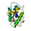
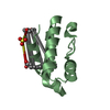
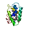
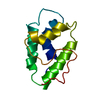
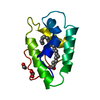
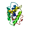
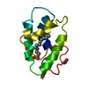
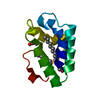
| Structure viewer | Molecule: MolmilJmol/JSmol |
|---|
- Downloads & links
-Download
| PDBx/mmCIF format | 1fk1.cif.gz | 27.9 KB | Display | PDBx/mmCIF format |
|---|---|---|---|---|
| PDB format | pdb1fk1.ent.gz | 18.1 KB | Display | PDB format |
| PDBx/mmJSON format | 1fk1.json.gz | Tree view | PDBx/mmJSON format | |
| Others |  Other downloads Other downloads |
-Validation report
| Arichive directory | https://data.pdbj.org/pub/pdb/validation_reports/fk/1fk1ftp://data.pdbj.org/pub/pdb/validation_reports/fk/1fk1 | HTTPS FTP |
|---|
-Related structure data
| Related structure data |  1fk0C  1fk2C  1fk3C  1fk4C  1fk5C  1fk6C  1fk7C  1mzlS S: Starting model for refinement C: citing same article ( |
|---|---|
| Similar structure data |

-Links
 PDBj
PDBj

- Assembly
| Deposited unit | 
| ||||||||
|---|---|---|---|---|---|---|---|---|---|
| 1 |
| ||||||||
| Unit cell |
|
-Components
| #1: Protein | Mass: 9062.161 Da / Num. of mol.: 1 / Source method: isolated from a natural source / Source: (natural) | ||||
|---|---|---|---|---|---|
| #2: Chemical | ChemComp-DAO /   Mass: 200.318 Da / Num. of mol.: 1 / Source method: obtained synthetically / Formula: C12H24O2 Mass: 200.318 Da / Num. of mol.: 1 / Source method: obtained synthetically / Formula: C12H24O2 | ||||
| #3: Chemical |   Mass: 46.025 Da / Num. of mol.: 3 / Source method: obtained synthetically / Formula: CH2O2 Mass: 46.025 Da / Num. of mol.: 3 / Source method: obtained synthetically / Formula: CH2O2#4: Water | ChemComp-HOH / |  Mass: 18.015 Da / Num. of mol.: 52 / Source method: isolated from a natural source / Formula: H2O Mass: 18.015 Da / Num. of mol.: 52 / Source method: isolated from a natural source / Formula: H2OHas protein modification | Y | |
-Experimental details
-Experiment
| Experiment | Method: X-RAY DIFFRACTION / Number of used crystals: 1 |
|---|
- Sample preparation
| Crystal | Density Matthews: 2.37 Å3/Da / Density % sol: 48.02 % | |||||||||||||||
|---|---|---|---|---|---|---|---|---|---|---|---|---|---|---|---|---|
| Crystal grow | Temperature: 298 K / Method: vapor diffusion, hanging drop Details: 4.2M Na formate, 0.2M potassium cyanide, VAPOR DIFFUSION, HANGING DROP, temperature 298.0K | |||||||||||||||
| Crystal grow | *PLUS Temperature: 24.5-25.5 ℃ | |||||||||||||||
| Components of the solutions | *PLUS
|
-Data collection
| Diffraction | Mean temperature: 298 K |
|---|---|
| Diffraction source | Source: ROTATING ANODE / Type: RIGAKU RU200 / Wavelength: 1.5418 |
| Detector | Type: ENRAF-NONIUS / Detector: AREA DETECTOR / Date: Sep 5, 1999 |
| Radiation | Protocol: SINGLE WAVELENGTH / Monochromatic (M) / Laue (L): M / Scattering type: x-ray |
| Radiation wavelength | Wavelength: 1.5418 Å / Relative weight: 1 |
| Reflection | Highest resolution: 1.76 Å / Num. obs: 8622 / % possible obs: 91.4 % / Redundancy: 2.69 % / Rmerge(I) obs: 0.044 |
| Reflection | *PLUS Num. obs: 8570 / Num. measured all: 23095 |
- Processing
| Software |
| ||||||||||||||||||||||||
|---|---|---|---|---|---|---|---|---|---|---|---|---|---|---|---|---|---|---|---|---|---|---|---|---|---|
| Refinement | Method to determine structure: molecular replacement Starting model: (PDB code:1MZL) Resolution: 1.8→8 Å / σ(F): 2
| ||||||||||||||||||||||||
| Refinement step | Cycle: LAST / Resolution: 1.8→8 Å
| ||||||||||||||||||||||||
| Refine LS restraints |
| ||||||||||||||||||||||||
| Software | *PLUS Name: X-PLOR / Version: 3.843 / Classification: refinement | ||||||||||||||||||||||||
| Refinement | *PLUS Highest resolution: 1.8 Å / Lowest resolution: 8 Å / σ(F): 2 / Rfactor obs: 0.195 / Rfactor Rfree: 0.211 | ||||||||||||||||||||||||
| Solvent computation | *PLUS | ||||||||||||||||||||||||
| Displacement parameters | *PLUS |
