 Movie
Movie Controller

[English] 日本語
 Yorodumi
- PDB-1fjh: THE CRYSTAL STRUCTURE OF 3-ALPHA-HYDROXYSTEROID DEHYDROGENASE FRO... -
+ Open data

- Basic information
| Entry | Database: PDB / ID: 1fjh | ||||||
|---|---|---|---|---|---|---|---|
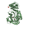
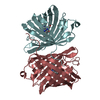
| Title | THE CRYSTAL STRUCTURE OF 3-ALPHA-HYDROXYSTEROID DEHYDROGENASE FROM COMAMONAS TESTOSTERONI, A MEMBER OF THE SHORT CHAIN DEHYDROGENASE/REDUCTASE FAMILY | ||||||
 Components Components | 3ALPHA-HYDROXYSTEROID DEHYDROGENASE/CARBONYL REDUCTASE | ||||||
 Keywords Keywords | OXIDOREDUCTASE / Short Chain Dehydrogenase / SDR / Carbonyl Reductase / Steroid / Hydroxysteroid / Xenobiotic / Metyrapone / Oligomerisation / Comamonas testosteroni | ||||||
| Function / homology |  Function and homology information Function and homology information3alpha-hydroxysteroid 3-dehydrogenase (Si-specific) / 3-alpha-hydroxysteroid 3-dehydrogenase [NAD(P)+] activity / nucleotide binding / cytoplasm Similarity search - Function | ||||||
| Biological species |  Comamonas testosteroni (bacteria) Comamonas testosteroni (bacteria) | ||||||
| Method |  X-RAY DIFFRACTION / Resolution: 1.68 Å X-RAY DIFFRACTION / Resolution: 1.68 Å | ||||||
 Authors Authors | Grimm, C. / Ficner, R. / Maser, E. / Klebe, G. / Reuter, K. | ||||||
 Citation Citation | Journal: J.Biol.Chem. / Year: 2000 Title: The crystal structure of 3alpha -hydroxysteroid dehydrogenase/carbonyl reductase from Comamonas testosteroni shows a novel oligomerization pattern within the short chain dehydrogenase/reductase family. Authors: Grimm, C. / Maser, E. / Mobus, E. / Klebe, G. / Reuter, K. / Ficner, R. | ||||||
| History |
|
- Structure visualization
| Structure viewer | Molecule: MolmilJmol/JSmol |
|---|
- Downloads & links
-Download
| PDBx/mmCIF format | 1fjh.cif.gz | 105 KB | Display | PDBx/mmCIF format |
|---|---|---|---|---|
| PDB format | pdb1fjh.ent.gz | 79.1 KB | Display | PDB format |
| PDBx/mmJSON format | 1fjh.json.gz | Tree view | PDBx/mmJSON format | |
| Others |  Other downloads Other downloads |
-Validation report
| Arichive directory | https://data.pdbj.org/pub/pdb/validation_reports/fj/1fjhftp://data.pdbj.org/pub/pdb/validation_reports/fj/1fjh | HTTPS FTP |
|---|
-Related structure data


-Links
 PDBj
PDBj


- Assembly
| Deposited unit | 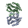
| ||||||||
|---|---|---|---|---|---|---|---|---|---|
| 1 |
| ||||||||
| Unit cell |
| ||||||||
| Details | The biological assembly is a dimer constructed from chain A by the two-fold NCS axis. |
-Components
| #1: Protein | Mass: 26420.430 Da / Num. of mol.: 2 / Source method: isolated from a natural source / Source: (natural) Comamonas testosteroni (bacteria)References: UniProt: Q9ZFY9, UniProt: P80702*PLUS, 3alpha-hydroxysteroid 3-dehydrogenase (Si-specific) #2: Water | ChemComp-HOH / |  Mass: 18.015 Da / Num. of mol.: 405 / Source method: isolated from a natural source / Formula: H2O Mass: 18.015 Da / Num. of mol.: 405 / Source method: isolated from a natural source / Formula: H2O |
|---|
-Experimental details
-Experiment
| Experiment | Method: X-RAY DIFFRACTION / Number of used crystals: 1 |
|---|
- Sample preparation
| Crystal | Density Matthews: 2.19 Å3/Da / Density % sol: 43.96 % | |||||||||||||||||||||||||||||||||||
|---|---|---|---|---|---|---|---|---|---|---|---|---|---|---|---|---|---|---|---|---|---|---|---|---|---|---|---|---|---|---|---|---|---|---|---|---|
| Crystal grow | Temperature: 277 K / Method: vapor diffusion, hanging drop / pH: 6.5 Details: PEG4000, ammonium acetate, cacodylate, pH 6.5, VAPOR DIFFUSION, HANGING DROP, temperature 277.0K | |||||||||||||||||||||||||||||||||||
| Crystal grow | *PLUS Temperature: 277 K / pH: 7.5 / Details: used macroseeding | |||||||||||||||||||||||||||||||||||
| Components of the solutions | *PLUS
|
-Data collection
| Diffraction | Mean temperature: 100 K |
|---|---|
| Diffraction source | Source: ROTATING ANODE / Type: RIGAKU / Wavelength: 1.5418 |
| Detector | Type: RIGAKU RAXIS / Detector: IMAGE PLATE / Date: Oct 20, 1999 |
| Radiation | Protocol: SINGLE WAVELENGTH / Monochromatic (M) / Laue (L): M / Scattering type: x-ray |
| Radiation wavelength | Wavelength: 1.5418 Å / Relative weight: 1 |
| Reflection | Resolution: 1.65→30.02 Å / Num. all: 50140 / Num. obs: 50140 / % possible obs: 97.7 % / Observed criterion σ(F): 0 / Observed criterion σ(I): 0 / Redundancy: 7.46 % / Biso Wilson estimate: 17.7 Å2 / Rmerge(I) obs: 0.043 / Net I/σ(I): 69.7 |
| Reflection shell | Resolution: 1.65→1.66 Å / Redundancy: 4.2 % / Rmerge(I) obs: 0.271 / % possible all: 74 |
| Reflection | *PLUS Num. measured all: 374072 |
| Reflection shell | *PLUS % possible obs: 74 % |
- Processing
| Software |
| |||||||||||||||||||||||||
|---|---|---|---|---|---|---|---|---|---|---|---|---|---|---|---|---|---|---|---|---|---|---|---|---|---|---|
| Refinement | Resolution: 1.68→30.02 Å / Rfactor Rfree error: 0.005 / Data cutoff high absF: 548597.76 / Data cutoff low absF: 0 / Isotropic thermal model: RESTRAINED / Cross valid method: THROUGHOUT / σ(F): 0 / σ(I): 2 / Stereochemistry target values: Engh & Huber
| |||||||||||||||||||||||||
| Solvent computation | Solvent model: FLAT MODEL / Bsol: 45.62 Å2 / ksol: 0.339 e/Å3 | |||||||||||||||||||||||||
| Displacement parameters | Biso mean: 20.2 Å2
| |||||||||||||||||||||||||
| Refine analyze |
| |||||||||||||||||||||||||
| Refinement step | Cycle: LAST / Resolution: 1.68→30.02 Å
| |||||||||||||||||||||||||
| Refine LS restraints |
| |||||||||||||||||||||||||
| LS refinement shell | Resolution: 1.68→1.79 Å / Rfactor Rfree error: 0.017 / Total num. of bins used: 6
| |||||||||||||||||||||||||
| Xplor file |
| |||||||||||||||||||||||||
| Software | *PLUS Name: CNS / Version: 1 / Classification: refinement | |||||||||||||||||||||||||
| Refinement | *PLUS σ(F): 0 / % reflection Rfree: 4 % / Rfactor obs: 0.2 / Rfactor Rwork: 0.2 | |||||||||||||||||||||||||
| Solvent computation | *PLUS | |||||||||||||||||||||||||
| Displacement parameters | *PLUS Biso mean: 20.2 Å2 | |||||||||||||||||||||||||
| Refine LS restraints | *PLUS
| |||||||||||||||||||||||||
| LS refinement shell | *PLUS Rfactor Rfree: 0.289 / % reflection Rfree: 3.9 % / Rfactor Rwork: 0.256 |
