 Movie
Movie Controller

[English] 日本語
 Yorodumi
- PDB-1fhl: CRYSTAL STRUCTURE OF BETA-1,4-GALACTANASE FROM ASPERGILLUS ACULEA... -
+ Open data

- Basic information
| Entry | Database: PDB / ID: 1fhl | ||||||
|---|---|---|---|---|---|---|---|
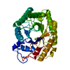
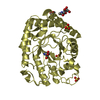
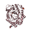
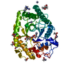
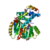
| Title | CRYSTAL STRUCTURE OF BETA-1,4-GALACTANASE FROM ASPERGILLUS ACULEATUS AT 293K | ||||||
 Components Components | BETA-1,4-GALACTANASE | ||||||
 Keywords Keywords | HYDROLASE / B/A barrel / glycosyl hydrolase / family 53 / clan GH-A | ||||||
| Function / homology |  Function and homology information Function and homology informationarabinogalactan endo-beta-1,4-galactanase / arabinogalactan endo-1,4-beta-galactosidase activity / glucosidase activity / pectin catabolic process Similarity search - Function | ||||||
| Biological species |  | ||||||
| Method |  X-RAY DIFFRACTION / Resolution: 2.3 Å X-RAY DIFFRACTION / Resolution: 2.3 Å | ||||||
 Authors Authors | Ryttersgaard, C. / Larsen, S. | ||||||
 Citation Citation | Journal: Biochemistry / Year: 2002 Title: Aspergillus aculeatus beta-1,4-Galactanase: Substrate Recognition and Relations to Other Glycoside Hydrolases in Clan GH-A Authors: Ryttersgaard, C. / Leggio, L.L. / Coutinho, P.M. / Henrissat, B. / Larsen, S. #1: Journal: Acta Crystallogr.,Sect.D / Year: 1999Title: Crystallization and Preliminary X-ray Studies of beta-1,4-galactanase from Aspergillus aculeatus Authors: Ryttersgaard, C. / Poulsen, J. / Christgau, S. / Sandal, T. / Dalboge, H. / Larsen, S. | ||||||
| History |
|
- Structure visualization
| Structure viewer | Molecule: MolmilJmol/JSmol |
|---|
- Downloads & links
-Download
| PDBx/mmCIF format | 1fhl.cif.gz | 75.9 KB | Display | PDBx/mmCIF format |
|---|---|---|---|---|
| PDB format | pdb1fhl.ent.gz | 57.3 KB | Display | PDB format |
| PDBx/mmJSON format | 1fhl.json.gz | Tree view | PDBx/mmJSON format | |
| Others |  Other downloads Other downloads |
-Validation report
| Summary document | 1fhl_validation.pdf.gz | 425.4 KB | Display | wwPDB validaton report |
|---|---|---|---|---|
| Full document | 1fhl_full_validation.pdf.gz | 432.6 KB | Display | |
| Data in XML | 1fhl_validation.xml.gz | 14.4 KB | Display | |
| Data in CIF | 1fhl_validation.cif.gz | 19.3 KB | Display | |
| Arichive directory | https://data.pdbj.org/pub/pdb/validation_reports/fh/1fhlftp://data.pdbj.org/pub/pdb/validation_reports/fh/1fhl | HTTPS FTP |
-Related structure data


-Links
 PDBj
PDBj- Assembly
| Deposited unit | 
| ||||||||
|---|---|---|---|---|---|---|---|---|---|
| 1 |
| ||||||||
| Unit cell |
| ||||||||
| Components on special symmetry positions |
| ||||||||
| Details | The biological assembly is a monomer |
-Components
| #1: Protein | Mass: 36775.438 Da / Num. of mol.: 1 / Source method: isolated from a natural source / Source: (natural) References: UniProt: P48842, arabinogalactan endo-beta-1,4-galactanase |
|---|---|
| #2: Water | ChemComp-HOH /  Mass: 18.015 Da / Num. of mol.: 59 / Source method: isolated from a natural source / Formula: H2O Mass: 18.015 Da / Num. of mol.: 59 / Source method: isolated from a natural source / Formula: H2O |
| Has protein modification | Y |
-Experimental details
-Experiment
| Experiment | Method: X-RAY DIFFRACTION / Number of used crystals: 1 |
|---|
- Sample preparation
| Crystal | Density Matthews: 2.36 Å3/Da / Density % sol: 47.81 % | ||||||||||||||||||||||||||||
|---|---|---|---|---|---|---|---|---|---|---|---|---|---|---|---|---|---|---|---|---|---|---|---|---|---|---|---|---|---|
| Crystal grow | Temperature: 298 K / Method: vapor diffusion, hanging drop / pH: 7.5 Details: PEG 400, calcium chloride, sodium hepes, pH 7.5, VAPOR DIFFUSION, HANGING DROP, temperature 298K | ||||||||||||||||||||||||||||
| Crystal grow | *PLUS Method: vapor diffusion, hanging dropDetails: Ryttersgaard, C., (1999) Acta Crystallogr.,Sect.D, 55, 929. | ||||||||||||||||||||||||||||
| Components of the solutions | *PLUS
|
-Data collection
| Diffraction | Mean temperature: 293 K |
|---|---|
| Diffraction source | Source: ROTATING ANODE / Type: RIGAKU RU200 / Wavelength: 1.5418 |
| Detector | Type: RIGAKU RAXIS II / Detector: IMAGE PLATE / Date: Mar 14, 1997 |
| Radiation | Protocol: SINGLE WAVELENGTH / Monochromatic (M) / Laue (L): M / Scattering type: x-ray |
| Radiation wavelength | Wavelength: 1.5418 Å / Relative weight: 1 |
| Reflection | Resolution: 2.3→28.8 Å / Num. all: 15582 / Num. obs: 15582 / % possible obs: 97.5 % / Observed criterion σ(F): 0 / Observed criterion σ(I): 0 / Redundancy: 4.6 % / Biso Wilson estimate: 30.82 Å2 / Rmerge(I) obs: 0.096 / Net I/σ(I): 7.1 |
| Reflection shell | Resolution: 2.3→2.42 Å / Redundancy: 4.4 % / Rmerge(I) obs: 0.392 / Num. unique all: 2154 / % possible all: 95.6 |
| Reflection | *PLUS Highest resolution: 2.3 Å / Lowest resolution: 28.9 Å |
| Reflection shell | *PLUS % possible obs: 95.6 % / Mean I/σ(I) obs: 61.4 |
- Processing
| Software |
| |||||||||||||||||||||||||
|---|---|---|---|---|---|---|---|---|---|---|---|---|---|---|---|---|---|---|---|---|---|---|---|---|---|---|
| Refinement | Resolution: 2.3→28.8 Å / σ(F): 0 / σ(I): 0 / Stereochemistry target values: Engh & Huber
| |||||||||||||||||||||||||
| Refinement step | Cycle: LAST / Resolution: 2.3→28.8 Å
| |||||||||||||||||||||||||
| Refine LS restraints |
| |||||||||||||||||||||||||
| Refinement | *PLUS Highest resolution: 2.3 Å / Lowest resolution: 28.9 Å | |||||||||||||||||||||||||
| Solvent computation | *PLUS | |||||||||||||||||||||||||
| Displacement parameters | *PLUS | |||||||||||||||||||||||||
| Refine LS restraints | *PLUS
|
