 Movie
Movie Controller

[English] 日本語
 Yorodumi
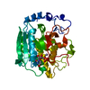
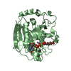
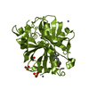
- PDB-1fg5: CRYSTAL STRUCTURE OF BOVINE ALPHA-1,3-GALACTOSYLTRANSFERASE CATAL... -
+ Open data

- Basic information
| Entry | Database: PDB / ID: 1fg5 | ||||||
|---|---|---|---|---|---|---|---|
| Title | CRYSTAL STRUCTURE OF BOVINE ALPHA-1,3-GALACTOSYLTRANSFERASE CATALYTIC DOMAIN. | ||||||
 Components Components | N-ACETYLLACTOSAMINIDE ALPHA-1,3-GALACTOSYLTRANSFERASE | ||||||
 Keywords Keywords | TRANSFERASE / ALPHA BETA ALPHA PROTEIN / NUCLEOTIDE BINDING PROTEIN / ROSSMANN FOLD | ||||||
| Function / homology |  Function and homology information Function and homology informationN-acetyllactosaminide 3-alpha-galactosyltransferase / N-acetyllactosaminide 3-alpha-galactosyltransferase activity / Golgi cisterna / : / Golgi cisterna membrane / : / glycosyltransferase activity / vesicle / carbohydrate metabolic process / Golgi apparatus / metal ion binding Similarity search - Function | ||||||
| Biological species |  | ||||||
| Method |  X-RAY DIFFRACTION / SYNCHROTRON / MAD / Resolution: 2.8 Å X-RAY DIFFRACTION / SYNCHROTRON / MAD / Resolution: 2.8 Å | ||||||
 Authors Authors | Gastinel, L.N. / Bignon, C. / Shaper, J.H. / Joziasse, D.H. | ||||||
 Citation Citation | Journal: EMBO J. / Year: 2001 Title: Bovine alpha1,3-galactosyltransferase catalytic domain structure and its relationship with ABO histo-blood group and glycosphingolipid glycosyltransferases. Authors: Gastinel, L.N. / Bignon, C. / Misra, A.K. / Hindsgaul, O. / Shaper, J.H. / Joziasse, D.H. | ||||||
| History |
|
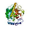
- Structure visualization
| Structure viewer | Molecule: MolmilJmol/JSmol |
|---|
- Downloads & links
-Download
| PDBx/mmCIF format | 1fg5.cif.gz | 71.8 KB | Display | PDBx/mmCIF format |
|---|---|---|---|---|
| PDB format | pdb1fg5.ent.gz | 53.8 KB | Display | PDB format |
| PDBx/mmJSON format | 1fg5.json.gz | Tree view | PDBx/mmJSON format | |
| Others |  Other downloads Other downloads |
-Validation report
| Summary document | 1fg5_validation.pdf.gz | 427.3 KB | Display | wwPDB validaton report |
|---|---|---|---|---|
| Full document | 1fg5_full_validation.pdf.gz | 447.2 KB | Display | |
| Data in XML | 1fg5_validation.xml.gz | 16.4 KB | Display | |
| Data in CIF | 1fg5_validation.cif.gz | 21 KB | Display | |
| Arichive directory | https://data.pdbj.org/pub/pdb/validation_reports/fg/1fg5ftp://data.pdbj.org/pub/pdb/validation_reports/fg/1fg5 | HTTPS FTP |
-Related structure data


-Links
 PDBj
PDBj

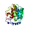
- Assembly
| Deposited unit | 
| ||||||||
|---|---|---|---|---|---|---|---|---|---|
| 1 |
| ||||||||
| Unit cell |
|
-Components
| #1: Protein | Mass: 36908.566 Da / Num. of mol.: 1 / Fragment: CATALYTIC DOMAIN Source method: isolated from a genetically manipulated source Source: (gene. exp.)  |
|---|---|
| #2: Water | ChemComp-HOH /  Mass: 18.015 Da / Num. of mol.: 43 / Source method: isolated from a natural source / Formula: H2O Mass: 18.015 Da / Num. of mol.: 43 / Source method: isolated from a natural source / Formula: H2O |
| Has protein modification | Y |
-Experimental details
-Experiment
| Experiment | Method: X-RAY DIFFRACTION / Number of used crystals: 1 |
|---|
- Sample preparation
| Crystal | Density Matthews: 3.5 Å3/Da / Density % sol: 64.85 % / Description: 0.9796 peak, 0.9800 inflection, 0.9324 remote | ||||||||||||||||||||||||||||||||||||||||||||||||
|---|---|---|---|---|---|---|---|---|---|---|---|---|---|---|---|---|---|---|---|---|---|---|---|---|---|---|---|---|---|---|---|---|---|---|---|---|---|---|---|---|---|---|---|---|---|---|---|---|---|
| Crystal grow | Temperature: 293 K / Method: vapor diffusion, hanging drop / pH: 6.5 Details: cacodylate, magnesium chloride, sodium acetate, pH 6.5, VAPOR DIFFUSION, HANGING DROP, temperature 293K | ||||||||||||||||||||||||||||||||||||||||||||||||
| Crystal grow | *PLUS Temperature: 20 ℃ / pH: 8 | ||||||||||||||||||||||||||||||||||||||||||||||||
| Components of the solutions | *PLUS
|
-Data collection
| Diffraction | Mean temperature: 100 K | ||||||||||||
|---|---|---|---|---|---|---|---|---|---|---|---|---|---|
| Diffraction source | Source: SYNCHROTRON / Site: ESRF  / Beamline: ID14-4 / Wavelength: 0.9796, 0.9800, 0.9324 / Beamline: ID14-4 / Wavelength: 0.9796, 0.9800, 0.9324 | ||||||||||||
| Detector | Type: ADSC QUANTUM / Detector: CCD / Date: Sep 15, 1999 | ||||||||||||
| Radiation | Protocol: MAD / Monochromatic (M) / Laue (L): M / Scattering type: x-ray | ||||||||||||
| Radiation wavelength |
| ||||||||||||
| Reflection | Resolution: 2.8→36.5 Å / Num. all: 42677 / Num. obs: 11867 / % possible obs: 95.7 % / Observed criterion σ(F): 2 / Observed criterion σ(I): 2 / Redundancy: 3.5 % / Biso Wilson estimate: 27 Å2 / Rmerge(I) obs: 0.075 / Net I/σ(I): 5.6 | ||||||||||||
| Reflection shell | Resolution: 2.8→2.95 Å / Redundancy: 3.4 % / Rmerge(I) obs: 0.12 / % possible all: 95.7 | ||||||||||||
| Reflection shell | *PLUS % possible obs: 95.7 % |
- Processing
| Software |
| |||||||||||||||||||||||||
|---|---|---|---|---|---|---|---|---|---|---|---|---|---|---|---|---|---|---|---|---|---|---|---|---|---|---|
| Refinement | Method to determine structure: MAD / Resolution: 2.8→36.5 Å / σ(F): 0 / σ(I): 0 / Stereochemistry target values: CNS, Brunger et al Details: maximum likelihood target using amplitudes and phase probability distribution
| |||||||||||||||||||||||||
| Refinement step | Cycle: LAST / Resolution: 2.8→36.5 Å
| |||||||||||||||||||||||||
| Refine LS restraints |
| |||||||||||||||||||||||||
| Software | *PLUS Name: CNS / Version: 0.9 / Classification: refinement | |||||||||||||||||||||||||
| Refinement | *PLUS Highest resolution: 2.8 Å / Lowest resolution: 36.5 Å / σ(F): 0 / Rfactor obs: 0.237 | |||||||||||||||||||||||||
| Solvent computation | *PLUS | |||||||||||||||||||||||||
| Displacement parameters | *PLUS | |||||||||||||||||||||||||
| Refine LS restraints | *PLUS Type: c_angle_deg / Dev ideal: 1.4 |
