 Movie
Movie Controller


+ Open data

- Basic information
| Entry | Database: PDB / ID: 1few | ||||||
|---|---|---|---|---|---|---|---|
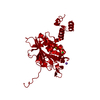
| Title | CRYSTAL STRUCTURE OF SMAC/DIABLO | ||||||
 Components Components | SECOND MITOCHONDRIA-DERIVED ACTIVATOR OF CASPASES | ||||||
 Keywords Keywords | APOPTOSIS / Smac / DIABLO / caspase activation / IAP inhibition | ||||||
| Function / homology |  Function and homology information Function and homology informationRelease of apoptotic factors from the mitochondria / CD40 receptor complex / SMAC, XIAP-regulated apoptotic response / Regulation of the apoptosome activity / SMAC (DIABLO) binds to IAPs / SMAC(DIABLO)-mediated dissociation of IAP:caspase complexes / intrinsic apoptotic signaling pathway in response to oxidative stress / extrinsic apoptotic signaling pathway via death domain receptors / intrinsic apoptotic signaling pathway / apoptotic signaling pathway ...Release of apoptotic factors from the mitochondria / CD40 receptor complex / SMAC, XIAP-regulated apoptotic response / Regulation of the apoptosome activity / SMAC (DIABLO) binds to IAPs / SMAC(DIABLO)-mediated dissociation of IAP:caspase complexes / intrinsic apoptotic signaling pathway in response to oxidative stress / extrinsic apoptotic signaling pathway via death domain receptors / intrinsic apoptotic signaling pathway / apoptotic signaling pathway / mitochondrial intermembrane space / cytoplasmic side of plasma membrane / neuron apoptotic process / positive regulation of apoptotic process / apoptotic process / mitochondrion / cytosol Similarity search - Function | ||||||
| Biological species |  Homo sapiens (human) Homo sapiens (human) | ||||||
| Method |  X-RAY DIFFRACTION / Resolution: 2.2 Å X-RAY DIFFRACTION / Resolution: 2.2 Å | ||||||
 Authors Authors | Chai, J. / Shi, Y. | ||||||
 Citation Citation | Journal: Nature / Year: 2000 Title: Structural and biochemical basis of apoptotic activation by Smac/DIABLO. Authors: Chai, J. / Du, C. / Wu, J.W. / Kyin, S. / Wang, X. / Shi, Y. | ||||||
| History |
|
- Structure visualization
| Structure viewer | Molecule: MolmilJmol/JSmol |
|---|
- Downloads & links
-Download
| PDBx/mmCIF format | 1few.cif.gz | 44.1 KB | Display | PDBx/mmCIF format |
|---|---|---|---|---|
| PDB format | pdb1few.ent.gz | 32.5 KB | Display | PDB format |
| PDBx/mmJSON format | 1few.json.gz | Tree view | PDBx/mmJSON format | |
| Others |  Other downloads Other downloads |
-Validation report
| Summary document | 1few_validation.pdf.gz | 361.1 KB | Display | wwPDB validaton report |
|---|---|---|---|---|
| Full document | 1few_full_validation.pdf.gz | 365.4 KB | Display | |
| Data in XML | 1few_validation.xml.gz | 5 KB | Display | |
| Data in CIF | 1few_validation.cif.gz | 6.9 KB | Display | |
| Arichive directory | https://data.pdbj.org/pub/pdb/validation_reports/fe/1fewftp://data.pdbj.org/pub/pdb/validation_reports/fe/1few | HTTPS FTP |
-Related structure data
| Similar structure data |
|---|


-Links
 PDBj
PDBj

- Assembly
| Deposited unit | 
| ||||||||
|---|---|---|---|---|---|---|---|---|---|
| 1 | 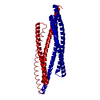
| ||||||||
| 2 | 
| ||||||||
| Unit cell |
| ||||||||
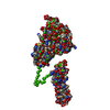
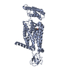
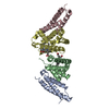
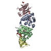
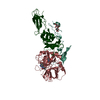
| Details | The biological assembly is a dimer constructed from chain A a symmetry partner generated by the two-fold. |
-Components
| #1: Protein | Mass: 20759.084 Da / Num. of mol.: 1 / Fragment: SMAC/DIABLO Source method: isolated from a genetically manipulated source Source: (gene. exp.) Homo sapiens (human) / Plasmid: PET-15B / Production host:  |
|---|
-Experimental details
-Experiment
| Experiment | Method: X-RAY DIFFRACTION / Number of used crystals: 1 |
|---|
- Sample preparation
| Crystal | Density Matthews: 2.88 Å3/Da / Density % sol: 57.3 % | |||||||||||||||||||||||||
|---|---|---|---|---|---|---|---|---|---|---|---|---|---|---|---|---|---|---|---|---|---|---|---|---|---|---|
| Crystal grow | Temperature: 276 K / Method: vapor diffusion, hanging drop / pH: 6.5 Details: Dioxane, Ammonium Sulfate, pH 6.5, VAPOR DIFFUSION, HANGING DROP, temperature 276.0K | |||||||||||||||||||||||||
| Crystal grow | *PLUS Temperature: 4 ℃ | |||||||||||||||||||||||||
| Components of the solutions | *PLUS
|
-Data collection
| Diffraction | Mean temperature: 100 K |
|---|---|
| Diffraction source | Source: ROTATING ANODE / Type: RIGAKU RU200 / Wavelength: 1.5418 |
| Detector | Type: RIGAKU RAXIS / Detector: IMAGE PLATE / Date: Jan 1, 2000 |
| Radiation | Protocol: SINGLE WAVELENGTH / Monochromatic (M) / Laue (L): M / Scattering type: x-ray |
| Radiation wavelength | Wavelength: 1.5418 Å / Relative weight: 1 |
| Reflection | Resolution: 2.2→99 Å / Num. all: 12907 / Num. obs: 12842 / % possible obs: 99.5 % / Observed criterion σ(F): 0 / Observed criterion σ(I): 0 / Redundancy: 6 % / Biso Wilson estimate: 37 Å2 / Rmerge(I) obs: 0.047 / Net I/σ(I): 16.3 |
| Reflection shell | Resolution: 2.2→2.28 Å / Redundancy: 2.5 % / Rmerge(I) obs: 0.242 / Num. unique all: 1219 / % possible all: 97.9 |
| Reflection | *PLUS Num. measured all: 97019 |
| Reflection shell | *PLUS % possible obs: 97.9 % |
- Processing
| Software |
| |||||||||||||||||||||||||
|---|---|---|---|---|---|---|---|---|---|---|---|---|---|---|---|---|---|---|---|---|---|---|---|---|---|---|
| Refinement | Resolution: 2.2→15 Å / σ(F): 2 / σ(I): 2 / Stereochemistry target values: Engh & Huber / Details: Used weighted full matrix least squares procedure.
| |||||||||||||||||||||||||
| Refinement step | Cycle: LAST / Resolution: 2.2→15 Å
| |||||||||||||||||||||||||
| Refine LS restraints |
| |||||||||||||||||||||||||
| Software | *PLUS Name: CNS / Classification: refinement | |||||||||||||||||||||||||
| Refinement | *PLUS Highest resolution: 2.2 Å / Lowest resolution: 15 Å / σ(F): 2 / % reflection Rfree: 5 % | |||||||||||||||||||||||||
| Solvent computation | *PLUS | |||||||||||||||||||||||||
| Displacement parameters | *PLUS |
