 Movie
Movie Controller

[English] 日本語
 Yorodumi
- PDB-1fer: STRUCTURE AT PH 6.5 OF FERREDOXIN I FROM AZOTOBACTER VINELANDII A... -
+ Open data

- Basic information
| Entry | Database: PDB / ID: 1fer | ||||||
|---|---|---|---|---|---|---|---|
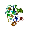
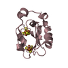
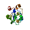
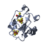
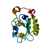
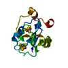
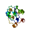
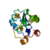
| Title | STRUCTURE AT PH 6.5 OF FERREDOXIN I FROM AZOTOBACTER VINELANDII AT 2.3 ANGSTROMS RESOLUTION | ||||||
 Components Components | FERREDOXIN I | ||||||
 Keywords Keywords | ELECTRON TRANSFER(IRON-SULFUR PROTEIN) | ||||||
| Function / homology |  Function and homology information Function and homology information3 iron, 4 sulfur cluster binding / 4 iron, 4 sulfur cluster binding / electron transfer activity / DNA binding / metal ion binding Similarity search - Function | ||||||
| Biological species |  Azotobacter vinelandii (bacteria) Azotobacter vinelandii (bacteria) | ||||||
| Method |  X-RAY DIFFRACTION / Resolution: 2.3 Å X-RAY DIFFRACTION / Resolution: 2.3 Å | ||||||
 Authors Authors | Merritt, E.A. / Stout, G.H. / Turley, S. / Sieker, L.C. / Jensen, L.H. / Orme-Johnson, W.H. | ||||||
 Citation Citation | Journal: Acta Crystallogr.,Sect.D / Year: 1993 Title: Structure at pH 6.5 of ferredoxin I from Azotobacter vinelandii at 2.3 A resolution. Authors: Merritt, E.A. / Stout, G.H. / Turley, S. / Sieker, L.C. / Jehsen, L.H. / Orme-Johnson, W.H. #1: Journal: Proc.Natl.Acad.Sci.USA / Year: 1988Title: Structure of Ferredoxin I from Azotobacter Vinelandii Authors: Stout, G.H. / Turley, S. / Sieker, L.C. / Jensen, L.H. | ||||||
| History |
| ||||||
| Remark 650 | HELIX HELIX ASSIGNMENTS WERE BASED ON ANALYSIS USING PROGRAM *DSSP* OF W. KABSCH AND C. SANDER. |
- Structure visualization
| Structure viewer | Molecule: MolmilJmol/JSmol |
|---|
- Downloads & links
-Download
| PDBx/mmCIF format | 1fer.cif.gz | 35.7 KB | Display | PDBx/mmCIF format |
|---|---|---|---|---|
| PDB format | pdb1fer.ent.gz | 24.4 KB | Display | PDB format |
| PDBx/mmJSON format | 1fer.json.gz | Tree view | PDBx/mmJSON format | |
| Others |  Other downloads Other downloads |
-Validation report
| Arichive directory | https://data.pdbj.org/pub/pdb/validation_reports/fe/1ferftp://data.pdbj.org/pub/pdb/validation_reports/fe/1fer | HTTPS FTP |
|---|
-Related structure data
| Similar structure data |
|---|

-Links
 PDBj
PDBj


- Assembly
| Deposited unit | 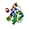
| ||||||||
|---|---|---|---|---|---|---|---|---|---|
| 1 |
| ||||||||
| Unit cell |
|
-Components
| #1: Protein | Mass: 12059.530 Da / Num. of mol.: 1 Source method: isolated from a genetically manipulated source Source: (gene. exp.) Azotobacter vinelandii (bacteria) / References: UniProt: P00214 |
|---|---|
| #2: Chemical | ChemComp-F3S /   Mass: 295.795 Da / Num. of mol.: 1 / Source method: obtained synthetically / Formula: Fe3S4 Mass: 295.795 Da / Num. of mol.: 1 / Source method: obtained synthetically / Formula: Fe3S4 |
| #3: Chemical | ChemComp-SF4 /   Mass: 351.640 Da / Num. of mol.: 1 / Source method: obtained synthetically / Formula: Fe4S4 Mass: 351.640 Da / Num. of mol.: 1 / Source method: obtained synthetically / Formula: Fe4S4 |
| #4: Water | ChemComp-HOH /  Mass: 18.015 Da / Num. of mol.: 98 / Source method: isolated from a natural source / Formula: H2O Mass: 18.015 Da / Num. of mol.: 98 / Source method: isolated from a natural source / Formula: H2O |
-Experimental details
-Experiment
| Experiment | Method: X-RAY DIFFRACTION |
|---|
- Sample preparation
| Crystal | Density Matthews: 3.06 Å3/Da / Density % sol: 59.77 % | ||||||||||||||||||||||||
|---|---|---|---|---|---|---|---|---|---|---|---|---|---|---|---|---|---|---|---|---|---|---|---|---|---|
| Crystal grow | *PLUS Temperature: 277 K / pH: 6.5 / Method: vapor diffusion, hanging drop | ||||||||||||||||||||||||
| Components of the solutions | *PLUS
|
-Data collection
| Radiation | Scattering type: x-ray |
|---|---|
| Radiation wavelength | Relative weight: 1 |
| Reflection | *PLUS Highest resolution: 2.3 Å / Lowest resolution: 30 Å / Num. obs: 7099 / Rmerge(I) obs: 3 |
- Processing
| Software |
| ||||||||||||
|---|---|---|---|---|---|---|---|---|---|---|---|---|---|
| Refinement | Rfactor Rwork: 0.17 / Highest resolution: 2.3 Å | ||||||||||||
| Refinement step | Cycle: LAST / Highest resolution: 2.3 Å
| ||||||||||||
| Refine LS restraints |
| ||||||||||||
| Refinement | *PLUS Highest resolution: 2.3 Å / Lowest resolution: 10 Å / Num. reflection obs: 6986 / Rfactor obs: 0.17 | ||||||||||||
| Solvent computation | *PLUS | ||||||||||||
| Displacement parameters | *PLUS Biso mean: 22.5 Å2 | ||||||||||||
| Refine LS restraints | *PLUS
|
