 Movie
Movie Controller

[English] 日本語
 Yorodumi
- PDB-1fd9: CRYSTAL STRUCTURE OF THE MACROPHAGE INFECTIVITY POTENTIATOR PROTE... -
+ Open data

- Basic information
| Entry | Database: PDB / ID: 1fd9 | ||||||
|---|---|---|---|---|---|---|---|
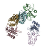
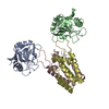
| Title | CRYSTAL STRUCTURE OF THE MACROPHAGE INFECTIVITY POTENTIATOR PROTEIN (MIP) A MAJOR VIRULENCE FACTOR FROM LEGIONELLA PNEUMOPHILA | ||||||
 Components Components | PROTEIN (MACROPHAGE INFECTIVITY POTENTIATOR PROTEIN) | ||||||
 Keywords Keywords | ISOMERASE / FKBP DOMAIN / LONG ALPHA HELIX / DIMERISATION VIA HELICAL INTERACTIONS | ||||||
| Function / homology |  Function and homology information Function and homology information: / peptidylprolyl isomerase / peptidyl-prolyl cis-trans isomerase activity / cell outer membrane / protein folding Similarity search - Function | ||||||
| Biological species |   Legionella pneumophila (bacteria) Legionella pneumophila (bacteria) | ||||||
| Method |  X-RAY DIFFRACTION / SYNCHROTRON / MAD / Resolution: 2.41 Å X-RAY DIFFRACTION / SYNCHROTRON / MAD / Resolution: 2.41 Å | ||||||
 Authors Authors | Riboldi-Tunnicliffe, A. / Jessen, S. / Konig, B. / Rahfeld, J. / Hacker, J. / Fischer, G. / Hilgenfeld, R. | ||||||
 Citation Citation | Journal: Nat.Struct.Biol. / Year: 2001 Title: Crystal structure of Mip, a prolylisomerase from Legionella pneumophila Authors: Riboldi-Tunnicliffe, A. / Konig, B. / Jessen, S. / Weiss, M.S. / Rahfeld, J. / Hacker, J. / Fischer, G. / Hilgenfeld, R. #1: Journal: Mol.Microbiol. / Year: 1992Title: Mip protein of Legionella pneumophila exhibits peptidyl-prolyl-cis/trans isomerase (PPlase) activity. Authors: Fischer, G. / Bang, H. / Ludwig, B. / Mann, K. / Hacker, J. #2: Journal: FEMS Microbiol.Rev. / Year: 1994Title: Characterization of Mip proteins of Legionella pneumophila. Authors: Ludwig, B. / Rahfeld, J. / Schmidt, B. / Mann, K. / Wintermeyer, E. / Fischer, G. / Hacker, J. #3: Journal: FEBS Lett. / Year: 1995Title: Small angle X-ray solution scattering study on the dimerization of the FKBP25mem from Legionella pneumophila Authors: Schmidt, B. / Konig, S. / Svergun, D. / Volkov, V. / Fischer, G. / Koch, M.H. | ||||||
| History |
|
- Structure visualization
| Structure viewer | Molecule: MolmilJmol/JSmol |
|---|
- Downloads & links
-Download
| PDBx/mmCIF format | 1fd9.cif.gz | 54 KB | Display | PDBx/mmCIF format |
|---|---|---|---|---|
| PDB format | pdb1fd9.ent.gz | 38.9 KB | Display | PDB format |
| PDBx/mmJSON format | 1fd9.json.gz | Tree view | PDBx/mmJSON format | |
| Others |  Other downloads Other downloads |
-Validation report
| Arichive directory | https://data.pdbj.org/pub/pdb/validation_reports/fd/1fd9ftp://data.pdbj.org/pub/pdb/validation_reports/fd/1fd9 | HTTPS FTP |
|---|
-Related structure data
| Similar structure data |
|---|


-Links
 PDBj
PDBj

- Assembly
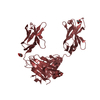
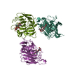
| Deposited unit | 
| ||||||||
|---|---|---|---|---|---|---|---|---|---|
| 1 | 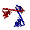
| ||||||||
| Unit cell |
|
-Components
| #1: Protein | Mass: 22869.012 Da / Num. of mol.: 1 Source method: isolated from a genetically manipulated source Source: (gene. exp.) Legionella pneumophila (bacteria) / Plasmid: PBLL106 / Production host: References: UniProt: P69059, UniProt: Q5ZXE0*PLUS, peptidylprolyl isomerase | ||
|---|---|---|---|
| #2: Chemical |   Mass: 65.409 Da / Num. of mol.: 2 / Source method: obtained synthetically / Formula: Zn Mass: 65.409 Da / Num. of mol.: 2 / Source method: obtained synthetically / Formula: Zn#3: Water | ChemComp-HOH / |  Mass: 18.015 Da / Num. of mol.: 96 / Source method: isolated from a natural source / Formula: H2O Mass: 18.015 Da / Num. of mol.: 96 / Source method: isolated from a natural source / Formula: H2O |
-Experimental details
-Experiment
| Experiment | Method: X-RAY DIFFRACTION / Number of used crystals: 1 |
|---|
- Sample preparation
| Crystal | Density Matthews: 3.65 Å3/Da / Density % sol: 65 % | ||||||||||||||||||||||||||||||||||||
|---|---|---|---|---|---|---|---|---|---|---|---|---|---|---|---|---|---|---|---|---|---|---|---|---|---|---|---|---|---|---|---|---|---|---|---|---|---|
| Crystal grow | Temperature: 288 K / Method: vapor diffusion, hanging drop / pH: 6.5 Details: PEG 8,000, zinc acetate, cacodylate, pH 6.5, VAPOR DIFFUSION, HANGING DROP, temperature 288K | ||||||||||||||||||||||||||||||||||||
| Crystal grow | *PLUS Method: vapor diffusion | ||||||||||||||||||||||||||||||||||||
| Components of the solutions | *PLUS
|
-Data collection
| Diffraction | Mean temperature: 100 K |
|---|---|
| Diffraction source | Source: SYNCHROTRON / Site: ELETTRA  / Beamline: 5.2R / Wavelength: 1.279 / Beamline: 5.2R / Wavelength: 1.279 |
| Detector | Type: MARRESEARCH / Detector: IMAGE PLATE / Date: Dec 2, 1998 |
| Radiation | Protocol: MAD / Monochromatic (M) / Laue (L): M / Scattering type: x-ray |
| Radiation wavelength | Wavelength: 1.279 Å / Relative weight: 1 |
| Reflection | Resolution: 2.41→22.4 Å / Num. obs: 13545 / % possible obs: 98.1 % / Observed criterion σ(I): 2 / Redundancy: 2.58 % / Biso Wilson estimate: 39.6 Å2 / Rmerge(I) obs: 0.072 / Rsym value: 0.075 / Net I/σ(I): 2.7 |
| Reflection shell | Resolution: 2.41→2.55 Å / Redundancy: 2.1 % / Rmerge(I) obs: 0.183 / Mean I/σ(I) obs: 2.4 / Rsym value: 0.23 / % possible all: 88.2 |
| Reflection | *PLUS Highest resolution: 2.41 Å / Lowest resolution: 22.4 Å / Num. obs: 10976 / % possible obs: 99.4 % / Redundancy: 4.8 % / Num. measured all: 52836 / Rmerge(I) obs: 0.103 |
| Reflection shell | *PLUS Highest resolution: 2.6 Å / Lowest resolution: 2.69 Å / % possible obs: 99.7 % / Rmerge(I) obs: 0.428 / Mean I/σ(I) obs: 3.2 |
- Processing
| Software |
| ||||||||||||||||||||||||||||||||||||||||||||||||||||||||||||||||||||||||||||||||
|---|---|---|---|---|---|---|---|---|---|---|---|---|---|---|---|---|---|---|---|---|---|---|---|---|---|---|---|---|---|---|---|---|---|---|---|---|---|---|---|---|---|---|---|---|---|---|---|---|---|---|---|---|---|---|---|---|---|---|---|---|---|---|---|---|---|---|---|---|---|---|---|---|---|---|---|---|---|---|---|---|---|
| Refinement | Method to determine structure: MAD / Resolution: 2.41→22.4 Å / Rfactor Rfree error: 0.009 / Cross valid method: THROUGHOUT / σ(F): 0 / Stereochemistry target values: ENGH & HUBER
| ||||||||||||||||||||||||||||||||||||||||||||||||||||||||||||||||||||||||||||||||
| Solvent computation | Solvent model: FLAT MODEL / Bsol: 84.82 Å2 / ksol: 0.37 e/Å3 | ||||||||||||||||||||||||||||||||||||||||||||||||||||||||||||||||||||||||||||||||
| Displacement parameters | Biso mean: 59.6 Å2
| ||||||||||||||||||||||||||||||||||||||||||||||||||||||||||||||||||||||||||||||||
| Refine analyze |
| ||||||||||||||||||||||||||||||||||||||||||||||||||||||||||||||||||||||||||||||||
| Refinement step | Cycle: LAST / Resolution: 2.41→22.4 Å
| ||||||||||||||||||||||||||||||||||||||||||||||||||||||||||||||||||||||||||||||||
| Refine LS restraints |
| ||||||||||||||||||||||||||||||||||||||||||||||||||||||||||||||||||||||||||||||||
| LS refinement shell | Resolution: 2.4→2.55 Å / Rfactor Rfree error: 0.024 / Total num. of bins used: 6
| ||||||||||||||||||||||||||||||||||||||||||||||||||||||||||||||||||||||||||||||||
| Xplor file |
| ||||||||||||||||||||||||||||||||||||||||||||||||||||||||||||||||||||||||||||||||
| Refinement | *PLUS Highest resolution: 2.41 Å / Lowest resolution: 22.4 Å / Num. reflection obs: 13169 / σ(F): 0 / Num. reflection Rfree: 1055 / % reflection Rfree: 8 % / Rfactor Rfree: 0.2805 | ||||||||||||||||||||||||||||||||||||||||||||||||||||||||||||||||||||||||||||||||
| Solvent computation | *PLUS | ||||||||||||||||||||||||||||||||||||||||||||||||||||||||||||||||||||||||||||||||
| Displacement parameters | *PLUS Biso mean: 59.6 Å2 | ||||||||||||||||||||||||||||||||||||||||||||||||||||||||||||||||||||||||||||||||
| Refine LS restraints | *PLUS
| ||||||||||||||||||||||||||||||||||||||||||||||||||||||||||||||||||||||||||||||||
| LS refinement shell | *PLUS Rfactor Rfree: 0.303 / % reflection Rfree: 8.1 % / Rfactor Rwork: 0.257 |
