 Movie
Movie Controller


+ Open data

- Basic information
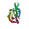
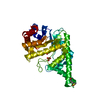
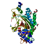
| Entry | Database: PDB / ID: 1fcu | ||||||
|---|---|---|---|---|---|---|---|
| Title | CRYSTAL STRUCTURE (TRIGONAL) OF BEE VENOM HYALURONIDASE | ||||||
 Components Components | HYALURONOGLUCOSAMINIDASE | ||||||
 Keywords Keywords | HYDROLASE / 7 stranded (beta/alpha) TIM barrel / allergen / glycosidase family 56 | ||||||
| Function / homology |  Function and homology information Function and homology informationhyaluronoglucosaminidase / hyalurononglucosaminidase activity / hyaluronan catabolic process / defense response / carbohydrate metabolic process / extracellular region Similarity search - Function | ||||||
| Biological species |  | ||||||
| Method |  X-RAY DIFFRACTION / SYNCHROTRON / Resolution: 2.1 Å X-RAY DIFFRACTION / SYNCHROTRON / Resolution: 2.1 Å | ||||||
 Authors Authors | Markovic-Housley, Z. / Miglierini, G. / Soldatova, L. / Rizkallah, P.J. / Mueller, U. / Schirmer, T. | ||||||
 Citation Citation | Journal: Structure Fold.Des. / Year: 2000 Title: Crystal structure of hyaluronidase, a major allergen of bee venom. Authors: Markovic-Housley, Z. / Miglierini, G. / Soldatova, L. / Rizkallah, P.J. / Muller, U. / Schirmer, T. | ||||||
| History |
|
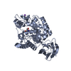
- Structure visualization
| Structure viewer | Molecule: MolmilJmol/JSmol |
|---|
- Downloads & links
-Download
| PDBx/mmCIF format | 1fcu.cif.gz | 84 KB | Display | PDBx/mmCIF format |
|---|---|---|---|---|
| PDB format | pdb1fcu.ent.gz | 62.9 KB | Display | PDB format |
| PDBx/mmJSON format | 1fcu.json.gz | Tree view | PDBx/mmJSON format | |
| Others |  Other downloads Other downloads |
-Validation report
| Summary document | 1fcu_validation.pdf.gz | 430.6 KB | Display | wwPDB validaton report |
|---|---|---|---|---|
| Full document | 1fcu_full_validation.pdf.gz | 437.8 KB | Display | |
| Data in XML | 1fcu_validation.xml.gz | 16.5 KB | Display | |
| Data in CIF | 1fcu_validation.cif.gz | 23.5 KB | Display | |
| Arichive directory | https://data.pdbj.org/pub/pdb/validation_reports/fc/1fcuftp://data.pdbj.org/pub/pdb/validation_reports/fc/1fcu | HTTPS FTP |
-Related structure data


-Links
 PDBj
PDBj- Assembly
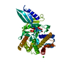
| Deposited unit | 
| ||||||||
|---|---|---|---|---|---|---|---|---|---|
| 1 |
| ||||||||
| Unit cell |
|
-Components
| #1: Protein | Mass: 40901.332 Da / Num. of mol.: 1 Source method: isolated from a genetically manipulated source Source: (gene. exp.) Trichoplusia ni (cabbage looper) / References: UniProt: Q08169, hyaluronoglucosaminidase |
|---|---|
| #2: Water | ChemComp-HOH /  Mass: 18.015 Da / Num. of mol.: 203 / Source method: isolated from a natural source / Formula: H2O Mass: 18.015 Da / Num. of mol.: 203 / Source method: isolated from a natural source / Formula: H2O |
| Has protein modification | Y |
-Experimental details
-Experiment
| Experiment | Method: X-RAY DIFFRACTION / Number of used crystals: 1 |
|---|
- Sample preparation
| Crystal | Density Matthews: 2.7 Å3/Da / Density % sol: 54 % | ||||||||||||||||||||||||||||||
|---|---|---|---|---|---|---|---|---|---|---|---|---|---|---|---|---|---|---|---|---|---|---|---|---|---|---|---|---|---|---|---|
| Crystal grow | Temperature: 298 K / Method: vapor diffusion, hanging drop / pH: 6.5 Details: PEG 8000, ammonium sulphate, Na cacodylate, pH 6.5, VAPOR DIFFUSION, HANGING DROP, temperature 298K | ||||||||||||||||||||||||||||||
| Crystal grow | *PLUS Temperature: 20 ℃ / pH: 5.4 | ||||||||||||||||||||||||||||||
| Components of the solutions | *PLUS
|
-Data collection
| Diffraction | Mean temperature: 293 K |
|---|---|
| Diffraction source | Source: SYNCHROTRON / Site: SRS  / Beamline: PX7.2 / Wavelength: 1.488 / Beamline: PX7.2 / Wavelength: 1.488 |
| Detector | Type: MARRESEARCH / Detector: IMAGE PLATE / Date: Mar 18, 1998 |
| Radiation | Protocol: SINGLE WAVELENGTH / Monochromatic (M) / Laue (L): M / Scattering type: x-ray |
| Radiation wavelength | Wavelength: 1.488 Å / Relative weight: 1 |
| Reflection | Resolution: 2.1→29.1 Å / Num. all: 23113 / Num. obs: 23113 / % possible obs: 87.5 % / Observed criterion σ(F): 0 / Observed criterion σ(I): 0 / Redundancy: 1.8 % / Biso Wilson estimate: 31.2 Å2 / Rmerge(I) obs: 0.111 / Net I/σ(I): 5 |
| Reflection shell | Resolution: 2.1→2.2 Å / Redundancy: 1.7 % / Rmerge(I) obs: 0.326 / % possible all: 62.5 |
| Reflection | *PLUS |
| Reflection shell | *PLUS % possible obs: 62.5 % / Mean I/σ(I) obs: 1.9 |
- Processing
| Software |
| ||||||||||||||||||||
|---|---|---|---|---|---|---|---|---|---|---|---|---|---|---|---|---|---|---|---|---|---|
| Refinement | Resolution: 2.1→8 Å / σ(F): 0 / σ(I): 0 / Stereochemistry target values: Engh & Huber
| ||||||||||||||||||||
| Refinement step | Cycle: LAST / Resolution: 2.1→8 Å
| ||||||||||||||||||||
| Refine LS restraints |
| ||||||||||||||||||||
| Software | *PLUS Name: REFMAC / Classification: refinement | ||||||||||||||||||||
| Refinement | *PLUS Highest resolution: 2.1 Å / Lowest resolution: 8 Å / σ(F): 0 / % reflection Rfree: 10 % / Rfactor Rwork: 0.2 | ||||||||||||||||||||
| Solvent computation | *PLUS | ||||||||||||||||||||
| Displacement parameters | *PLUS |
