 Movie
Movie Controller


+ Open data

- Basic information
| Entry | Database: PDB / ID: 1faj | ||||||
|---|---|---|---|---|---|---|---|
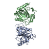
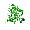
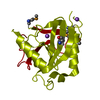
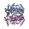
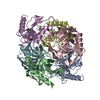
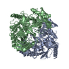
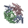
| Title | INORGANIC PYROPHOSPHATASE | ||||||
 Components Components | SOLUBLE INORGANIC PYROPHOSPHATASE | ||||||
 Keywords Keywords | INORGANIC PYROPHOSPHATASE / HYDROLASE / MAGNESIUM | ||||||
| Function / homology |  Function and homology information Function and homology informationinorganic triphosphate phosphatase activity / inorganic diphosphatase / inorganic diphosphate phosphatase activity / phosphate-containing compound metabolic process / magnesium ion binding / zinc ion binding / membrane / cytosol Similarity search - Function | ||||||
| Biological species |  | ||||||
| Method |  X-RAY DIFFRACTION / Resolution: 2.15 Å X-RAY DIFFRACTION / Resolution: 2.15 Å | ||||||
 Authors Authors | Kankare, J.A. / Salminen, T. / Goldman, A. | ||||||
 Citation Citation | Journal: Acta Crystallogr.,Sect.D / Year: 1996 Title: Structure of Escherichia coli inorganic pyrophosphatase at 2.2 A resolution. Authors: Kankare, J. / Salminen, T. / Lahti, R. / Cooperman, B.S. / Baykov, A.A. / Goldman, A. #1: Journal: Acta Crystallogr.,Sect.D / Year: 1995Title: New Crystal Forms of Escherichia Coli and Saccharomyces Cerevisiae Soluble Inorganic Pyrophosphatase Authors: Heikinheimo, P. / Salminen, T. / Cooperman, B. / Lahti, R. / Goldman, A. #2: Journal: Protein Eng. / Year: 1994Title: The Structure of E.Coli Soluble Inorganic Pyrophosphatase at 2.7 A Resolution Authors: Kankare, J. / Neal, G.S. / Salminen, T. / Glumhoff, T. / Cooperman, B.S. / Lahti, R. / Goldman, A. | ||||||
| History |
|
- Structure visualization
| Structure viewer | Molecule: MolmilJmol/JSmol |
|---|
- Downloads & links
-Download
| PDBx/mmCIF format | 1faj.cif.gz | 46.2 KB | Display | PDBx/mmCIF format |
|---|---|---|---|---|
| PDB format | pdb1faj.ent.gz | 33.4 KB | Display | PDB format |
| PDBx/mmJSON format | 1faj.json.gz | Tree view | PDBx/mmJSON format | |
| Others |  Other downloads Other downloads |
-Validation report
| Arichive directory | https://data.pdbj.org/pub/pdb/validation_reports/fa/1fajftp://data.pdbj.org/pub/pdb/validation_reports/fa/1faj | HTTPS FTP |
|---|
-Related structure data


-Links
 PDBj
PDBj- Assembly
| Deposited unit | 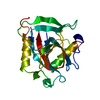
| ||||||||
|---|---|---|---|---|---|---|---|---|---|
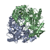
| 1 | x 6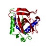
| ||||||||
| Unit cell |
| ||||||||
| Details | THE ACTIVE ENZYME IS A HEXAMER. THE ASYMMETRIC UNIT OF THIS CRYSTAL FORM CONTAINS A MONOMER OF THE ACTIVE HEXAMER. THE WHOLE HEXAMER CAN BE GENERATED AS FOLLOWS I) APPLY TWICE THE CRYSTALLOGRAPHIC THREE-FOLD ROTATION OPERATION AROUND C-AXIS TO GENERATE A TRIMER. II) ROTATE THE TRIMER AROUND THE CRYSTALLOGRAPHIC TWO-FOLD AND TRANSLATE (0. 0. 1.) IN FRACTIONAL UNITS. 1.5 B=111.5 C=76.5 |
-Components
| #1: Protein | Mass: 19597.334 Da / Num. of mol.: 1 / Source method: isolated from a natural source / Source: (natural) |
|---|---|
| #2: Water | ChemComp-HOH /  Mass: 18.015 Da / Num. of mol.: 81 / Source method: isolated from a natural source / Formula: H2O Mass: 18.015 Da / Num. of mol.: 81 / Source method: isolated from a natural source / Formula: H2O |
-Experimental details
-Experiment
| Experiment | Method: X-RAY DIFFRACTION |
|---|
- Sample preparation
| Crystal |
| ||||||||||||||||||||||||||||||||||||||||||
|---|---|---|---|---|---|---|---|---|---|---|---|---|---|---|---|---|---|---|---|---|---|---|---|---|---|---|---|---|---|---|---|---|---|---|---|---|---|---|---|---|---|---|---|
| Crystal grow | *PLUS pH: 8 / Method: vapor diffusion, hanging drop | ||||||||||||||||||||||||||||||||||||||||||
| Components of the solutions | *PLUS
|
-Data collection
| Diffraction source | Wavelength: 1.5418 |
|---|---|
| Detector | Type: RIGAKU RAXIS IIC / Detector: IMAGE PLATE |
| Radiation | Scattering type: x-ray |
| Radiation wavelength | Wavelength: 1.5418 Å / Relative weight: 1 |
| Reflection | Resolution: 2→20 Å / Num. obs: 12452 / % possible obs: 99.9 % / Observed criterion σ(I): 2 / Rmerge(I) obs: 0.073 |
| Reflection | *PLUS Num. measured all: 106709 |
| Reflection shell | *PLUS Highest resolution: 2 Å / Lowest resolution: 2.1 Å / % possible obs: 99.7 % / Mean I/σ(I) obs: 1.74 |
- Processing
| Software |
| ||||||||||||||||||||||||||||||||||||||||||||||||||||||||||||
|---|---|---|---|---|---|---|---|---|---|---|---|---|---|---|---|---|---|---|---|---|---|---|---|---|---|---|---|---|---|---|---|---|---|---|---|---|---|---|---|---|---|---|---|---|---|---|---|---|---|---|---|---|---|---|---|---|---|---|---|---|---|
| Refinement | Resolution: 2.15→8 Å / σ(F): 2
| ||||||||||||||||||||||||||||||||||||||||||||||||||||||||||||
| Displacement parameters | Biso mean: 33.7 Å2 | ||||||||||||||||||||||||||||||||||||||||||||||||||||||||||||
| Refinement step | Cycle: LAST / Resolution: 2.15→8 Å
| ||||||||||||||||||||||||||||||||||||||||||||||||||||||||||||
| Refine LS restraints |
| ||||||||||||||||||||||||||||||||||||||||||||||||||||||||||||
| Software | *PLUS Name: X-PLOR / Classification: refinement | ||||||||||||||||||||||||||||||||||||||||||||||||||||||||||||
| Refinement | *PLUS | ||||||||||||||||||||||||||||||||||||||||||||||||||||||||||||
| Solvent computation | *PLUS | ||||||||||||||||||||||||||||||||||||||||||||||||||||||||||||
| Displacement parameters | *PLUS | ||||||||||||||||||||||||||||||||||||||||||||||||||||||||||||
| Refine LS restraints | *PLUS
|
