 Movie
Movie Controller


+ Open data

- Basic information
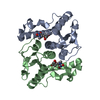
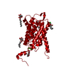
| Entry | Database: PDB / ID: 1f8x | ||||||
|---|---|---|---|---|---|---|---|
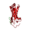
| Title | CRYSTAL STRUCTURE OF NUCLEOSIDE 2-DEOXYRIBOSYLTRANSFERASE | ||||||
 Components Components | NUCLEOSIDE 2-DEOXYRIBOSYLTRANSFERASE | ||||||
 Keywords Keywords | TRANSFERASE / active site / alpha/beta protein / biocatalyst / nucleoside | ||||||
| Function / homology |  Function and homology information Function and homology informationnucleoside deoxyribosyltransferase / nucleotide salvage / nucleoside deoxyribosyltransferase activity Similarity search - Function | ||||||
| Biological species |  Lactobacillus leichmannii (bacteria) Lactobacillus leichmannii (bacteria) | ||||||
| Method |  X-RAY DIFFRACTION / Resolution: 2.5 Å X-RAY DIFFRACTION / Resolution: 2.5 Å | ||||||
 Authors Authors | Armstrong, S.R. / Cook, W.J. / Short, S.A. / Ealick, S.E. | ||||||
 Citation Citation | Journal: Structure / Year: 1996 Title: Crystal structures of nucleoside 2-deoxyribosyltransferase in native and ligand-bound forms reveal architecture of the active site. Authors: Armstrong, S.R. / Cook, W.J. / Short, S.A. / Ealick, S.E. #1: Journal: J.Biol.Chem. / Year: 1990Title: Crystallization and Preliminary X-ray Investigation of Recombinant Lactobacillus leichmannii Nucleoside Deoxyribosyltransferase Authors: Cook, W.J. / Short, S.A. / Ealick, S.E. | ||||||
| History |
|
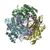
- Structure visualization
| Structure viewer | Molecule: MolmilJmol/JSmol |
|---|
- Downloads & links
-Download
| PDBx/mmCIF format | 1f8x.cif.gz | 73.8 KB | Display | PDBx/mmCIF format |
|---|---|---|---|---|
| PDB format | pdb1f8x.ent.gz | 56.8 KB | Display | PDB format |
| PDBx/mmJSON format | 1f8x.json.gz | Tree view | PDBx/mmJSON format | |
| Others |  Other downloads Other downloads |
-Validation report
| Arichive directory | https://data.pdbj.org/pub/pdb/validation_reports/f8/1f8xftp://data.pdbj.org/pub/pdb/validation_reports/f8/1f8x | HTTPS FTP |
|---|
-Related structure data


-Links
 PDBj
PDBj- Assembly
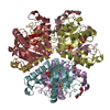
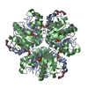
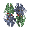
| Deposited unit | 
| ||||||||
|---|---|---|---|---|---|---|---|---|---|
| 1 | 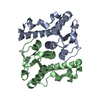
| ||||||||
| Unit cell |
|
-Components
| #1: Protein | Mass: 18098.354 Da / Num. of mol.: 2 / Source method: isolated from a natural source / Source: (natural) Lactobacillus leichmannii (bacteria)References: UniProt: Q9R5V5, nucleoside deoxyribosyltransferase #2: Water | ChemComp-HOH / |  Mass: 18.015 Da / Num. of mol.: 80 / Source method: isolated from a natural source / Formula: H2O Mass: 18.015 Da / Num. of mol.: 80 / Source method: isolated from a natural source / Formula: H2O |
|---|
-Experimental details
-Experiment
| Experiment | Method: X-RAY DIFFRACTION / Number of used crystals: 3 |
|---|
- Sample preparation
| Crystal | Density Matthews: 3.96 Å3/Da / Density % sol: 68.95 % | ||||||||||||||||||||||||||||||
|---|---|---|---|---|---|---|---|---|---|---|---|---|---|---|---|---|---|---|---|---|---|---|---|---|---|---|---|---|---|---|---|
| Crystal grow | Temperature: 296 K / Method: vapor diffusion, hanging drop / pH: 5.4 Details: ammonium sulfate, citrate buffer, pH 5.4, VAPOR DIFFUSION, HANGING DROP, temperature 296K | ||||||||||||||||||||||||||||||
| Crystal grow | *PLUS | ||||||||||||||||||||||||||||||
| Components of the solutions | *PLUS
|
-Data collection
| Diffraction |
| ||||||||||||||||
|---|---|---|---|---|---|---|---|---|---|---|---|---|---|---|---|---|---|
| Diffraction source |
| ||||||||||||||||
| Detector |
| ||||||||||||||||
| Radiation | Protocol: SINGLE WAVELENGTH / Monochromatic (M) / Laue (L): M / Scattering type: x-ray | ||||||||||||||||
| Radiation wavelength | Wavelength: 1.5418 Å / Relative weight: 1 | ||||||||||||||||
| Reflection | Resolution: 2.5→100 Å / Num. all: 223078 / Num. obs: 19875 / % possible obs: 95 % / Observed criterion σ(F): 0 / Observed criterion σ(I): 0 / Redundancy: 11.2 % / Rmerge(I) obs: 0.115 |
- Processing
| Software |
| ||||||||||||||||||||
|---|---|---|---|---|---|---|---|---|---|---|---|---|---|---|---|---|---|---|---|---|---|
| Refinement | Resolution: 2.5→5 Å / σ(F): 2 / σ(I): 0 / Stereochemistry target values: Engh & Huber
| ||||||||||||||||||||
| Refinement step | Cycle: LAST / Resolution: 2.5→5 Å
| ||||||||||||||||||||
| Refine LS restraints |
|
