 Movie
Movie Controller


+ Open data

- Basic information
| Entry | Database: PDB / ID: 1f0k | ||||||
|---|---|---|---|---|---|---|---|
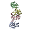
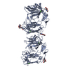
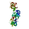
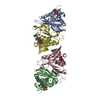
| Title | THE 1.9 ANGSTROM CRYSTAL STRUCTURE OF E. COLI MURG | ||||||
 Components Components | UDP-N-ACETYLGLUCOSAMINE-N-ACETYLMURAMYL-(PENTAPEPTIDE) PYROPHOSPHORYL-UNDECAPRENOL N-ACETYLGLUCOSAMINE TRANSFERASE | ||||||
 Keywords Keywords | TRANSFERASE / Rossmann fold | ||||||
| Function / homology |  Function and homology information Function and homology informationundecaprenyldiphospho-muramoylpentapeptide beta-N-acetylglucosaminyltransferase / undecaprenyldiphospho-muramoylpentapeptide beta-N-acetylglucosaminyltransferase activity / UDP-N-acetyl-D-glucosamine:N-acetylmuramoyl-L-alanyl-D-glutamyl-meso-2,6-diaminopimelyl-D-alanyl-D-alanine-diphosphoundecaprenol 4-beta-N-acetylglucosaminlytransferase activity / peptidoglycan biosynthetic process / cell wall organization / regulation of cell shape / carbohydrate metabolic process / cell division / plasma membrane Similarity search - Function | ||||||
| Biological species |  | ||||||
| Method |  X-RAY DIFFRACTION / SYNCHROTRON / Resolution: 1.9 Å X-RAY DIFFRACTION / SYNCHROTRON / Resolution: 1.9 Å | ||||||
 Authors Authors | Ha, S. / Walker, D. / Shi, Y. / Walker, S. | ||||||
 Citation Citation | Journal: Protein Sci. / Year: 2000 Title: The 1.9 A crystal structure of Escherichia coli MurG, a membrane-associated glycosyltransferase involved in peptidoglycan biosynthesis. Authors: Ha, S. / Walker, D. / Shi, Y. / Walker, S. | ||||||
| History |
|
- Structure visualization
| Structure viewer | Molecule: MolmilJmol/JSmol |
|---|
- Downloads & links
-Download
| PDBx/mmCIF format | 1f0k.cif.gz | 144.6 KB | Display | PDBx/mmCIF format |
|---|---|---|---|---|
| PDB format | pdb1f0k.ent.gz | 113.5 KB | Display | PDB format |
| PDBx/mmJSON format | 1f0k.json.gz | Tree view | PDBx/mmJSON format | |
| Others |  Other downloads Other downloads |
-Validation report
| Arichive directory | https://data.pdbj.org/pub/pdb/validation_reports/f0/1f0kftp://data.pdbj.org/pub/pdb/validation_reports/f0/1f0k | HTTPS FTP |
|---|
-Related structure data
| Similar structure data |
|---|


-Links
 PDBj
PDBj- Assembly
| Deposited unit | 
| ||||||||
|---|---|---|---|---|---|---|---|---|---|
| 1 |
| ||||||||
| Unit cell |
|
-Components
| #1: Protein | Mass: 39060.992 Da / Num. of mol.: 2 Source method: isolated from a genetically manipulated source Source: (gene. exp.) References: UniProt: P17443, Transferases; Glycosyltransferases; Hexosyltransferases #2: Chemical | ChemComp-SO4 / |   Mass: 96.063 Da / Num. of mol.: 1 / Source method: obtained synthetically / Formula: SO4 Mass: 96.063 Da / Num. of mol.: 1 / Source method: obtained synthetically / Formula: SO4#3: Water | ChemComp-HOH / |  Mass: 18.015 Da / Num. of mol.: 296 / Source method: isolated from a natural source / Formula: H2O Mass: 18.015 Da / Num. of mol.: 296 / Source method: isolated from a natural source / Formula: H2O |
|---|
-Experimental details
-Experiment
| Experiment | Method: X-RAY DIFFRACTION / Number of used crystals: 1 |
|---|
- Sample preparation
| Crystal | Density Matthews: 2.85 Å3/Da / Density % sol: 56.9 % | ||||||||||||||||||||||||||||||||||||||||||||||||||||||
|---|---|---|---|---|---|---|---|---|---|---|---|---|---|---|---|---|---|---|---|---|---|---|---|---|---|---|---|---|---|---|---|---|---|---|---|---|---|---|---|---|---|---|---|---|---|---|---|---|---|---|---|---|---|---|---|
| Crystal grow | Temperature: 298 K / Method: vapor diffusion, hanging drop / pH: 6.5 Details: NaMES, ammonium sulfate, TritonX-100, DTT, pH 6.5, VAPOR DIFFUSION, HANGING DROP, temperature 298.0K | ||||||||||||||||||||||||||||||||||||||||||||||||||||||
| Crystal grow | *PLUS pH: 7.9 Details: drop consists of equal volume of protein and reservoir solutions | ||||||||||||||||||||||||||||||||||||||||||||||||||||||
| Components of the solutions | *PLUS
|
-Data collection
| Diffraction | Mean temperature: 100 K |
|---|---|
| Diffraction source | Source: SYNCHROTRON / Site: APS  / Beamline: 14-BM-C / Wavelength: 1.0092 / Beamline: 14-BM-C / Wavelength: 1.0092 |
| Detector | Type: ADSC QUANTUM 4 / Detector: CCD / Date: Nov 11, 1999 |
| Radiation | Protocol: SINGLE WAVELENGTH / Monochromatic (M) / Laue (L): M / Scattering type: x-ray |
| Radiation wavelength | Wavelength: 1.0092 Å / Relative weight: 1 |
| Reflection | Resolution: 1.9→40 Å / Num. all: 65567 / Num. obs: 65567 / % possible obs: 97.7 % / Observed criterion σ(F): 1 / Observed criterion σ(I): 1 / Redundancy: 4.4 % / Biso Wilson estimate: 26 Å2 / Rmerge(I) obs: 0.032 / Net I/σ(I): 41.9 |
| Reflection shell | Resolution: 1.9→1.97 Å / Redundancy: 3.79 % / Rmerge(I) obs: 0.187 / Num. unique all: 6450 / % possible all: 96.4 |
| Reflection | *PLUS Num. measured all: 288150 |
| Reflection shell | *PLUS % possible obs: 96.4 % / Mean I/σ(I) obs: 7 |
- Processing
| Software |
| |||||||||||||||||||||||||
|---|---|---|---|---|---|---|---|---|---|---|---|---|---|---|---|---|---|---|---|---|---|---|---|---|---|---|
| Refinement | Resolution: 1.9→40 Å / σ(F): 2 / σ(I): 2
| |||||||||||||||||||||||||
| Refinement step | Cycle: LAST / Resolution: 1.9→40 Å
| |||||||||||||||||||||||||
| Refine LS restraints |
| |||||||||||||||||||||||||
| Software | *PLUS Name: 'CNS' / Classification: refinement | |||||||||||||||||||||||||
| Refinement | *PLUS Rfactor Rfree: 0.247 | |||||||||||||||||||||||||
| Solvent computation | *PLUS | |||||||||||||||||||||||||
| Displacement parameters | *PLUS |
