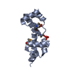
- PDB-1ezt: HIGH-RESOLUTION SOLUTION STRUCTURE OF FREE RGS4 BY NMR -
+
Open data
ID or keywords:
Loading...
-
Basic information
Entry
Database: PDB / ID: 1ezt
Title
HIGH-RESOLUTION SOLUTION STRUCTURE OF FREE RGS4 BY NMR
Components
REGULATOR OF G-PROTEIN SIGNALING 4
Keywords
SIGNALING PROTEIN INHIBITOR / 4-helix-bundle / free form of protein
Function / homology
Function and homology information
negative regulation of glycine import across plasma membrane / negative regulation of dopamine receptor signaling pathway / negative regulation of potassium ion transmembrane transport / dorsal root ganglion development / negative regulation of cell growth involved in cardiac muscle cell development / regulation of actin filament organization / regulation of potassium ion transmembrane transport / G alpha (q) signalling events / G alpha (i) signalling events / negative regulation of G protein-coupled receptor signaling pathway ...negative regulation of glycine import across plasma membrane / negative regulation of dopamine receptor signaling pathway / negative regulation of potassium ion transmembrane transport / dorsal root ganglion development / negative regulation of cell growth involved in cardiac muscle cell development / regulation of actin filament organization / regulation of potassium ion transmembrane transport / G alpha (q) signalling events / G alpha (i) signalling events / negative regulation of G protein-coupled receptor signaling pathway / regulation of calcium ion transport / G-protein alpha-subunit binding / positive regulation of heart rate / positive regulation of excitatory postsynaptic potential / GTPase activator activity / response to amphetamine / response to cocaine / response to ethanol / protein kinase binding / protein-containing complex / nucleus / cytoplasm Similarity search - Function
Regulator of G-protein signalling 4, RGS domain / Regulator of G-protein Signalling 4; domain 1 - #10 / Regulator of G-protein Signalling 4; domain 1 / RGS, subdomain 1/3 / Regulator of G-protein Signalling 4, domain 2 / Regulator of G-protein Signalling 4; domain 2 / Regulator of G protein signaling domain / RGS domain / RGS domain profile. / Regulator of G protein signalling domain ...Regulator of G-protein signalling 4, RGS domain / Regulator of G-protein Signalling 4; domain 1 - #10 / Regulator of G-protein Signalling 4; domain 1 / RGS, subdomain 1/3 / Regulator of G-protein Signalling 4, domain 2 / Regulator of G-protein Signalling 4; domain 2 / Regulator of G protein signaling domain / RGS domain / RGS domain profile. / Regulator of G protein signalling domain / RGS, subdomain 2 / RGS domain superfamily / Orthogonal Bundle / Mainly Alpha Similarity search - Domain/homology
Mass: 19145.711 Da / Num. of mol.: 1 / Fragment: CORE RGS DOMAIN Source method: isolated from a genetically manipulated source Details: SIX RESIDUE HISTIDINE TAG AT C-TERMINUS / Source: (gene. exp.) Rattus norvegicus (Norway rat) / Organ: BRAIN / Plasmid: PRGS4 / Production host: Escherichia coli (E. coli) / References: UniProt: P49799
-
Experimental details
-
Experiment
Experiment
Method: SOLUTION NMR
NMR experiment
Conditions-ID
Experiment-ID
Solution-ID
Type
1
1
2
3D 13C-separated NOESY
1
2
3
3D 15N-separated NOESY
1
3
3
HNHA
-
Sample preparation
Details
Solution-ID
Contents
Solvent system
1
1 mM RGS4 U-15N,13C; 50mM phosphate buffer, 2 mM NaN3, 50 mM deuterated DTT, pH 6.0
90% H2O/10% D2O
2
1 mM RGS4 U-15N,13C; 50mM phosphate buffer, 2 mM NaN3, 50 mM deuterated DTT, pH 6.0
100% D2O
3
1 mM RGS4 U-15N; 50mM phosphate buffer, 2 mM NaN3, 50 mM deuterated DTT, pH 6.0
90% H2O/10% D2O
Sample conditions
Ionic strength: 50 mM K / pH: 6 / Pressure: ambient / Temperature: 303 K
Crystal grow
*PLUS
Method: other / Details: NMR
-
NMR measurement
NMR spectrometer
Type: Bruker AMX / Manufacturer: Bruker / Model: AMX / Field strength: 600 MHz
-
Processing
NMR software
Name
Version
Developer
Classification
X-PLOR
3.84
Brunger
structuresolution
NMRPipe
1.7
Delaglio
processing
PIPP
4.2.8
Garrett
dataanalysis
XwinNMR
2
Bruker
collection
X-PLOR
3.84
Brunger
refinement
Refinement
Method: distance geometry simulated annealing / Software ordinal: 1 Details: The structure is based on a total of 2871 restraints, 1960 approximate interproton distance restraints, 78 distance restraints for 39 backbone hydrogen bonds, 431 torsion angle restraints, ...Details: The structure is based on a total of 2871 restraints, 1960 approximate interproton distance restraints, 78 distance restraints for 39 backbone hydrogen bonds, 431 torsion angle restraints, 132 3JHNa restraints, 136 Ca and 134 Cb chemical shift restraints. Structure also refined with conformational database target function.
NMR ensemble
Conformers submitted total number: 1
+
About Yorodumi
-
News
-
Feb 9, 2022. New format data for meta-information of EMDB entries
New format data for meta-information of EMDB entries
Version 3 of the EMDB header file is now the official format.
The previous official version 1.9 will be removed from the archive.
In the structure databanks used in Yorodumi, some data are registered as the other names, "COVID-19 virus" and "2019-nCoV". Here are the details of the virus and the list of structure data.
Jan 31, 2019. EMDB accession codes are about to change! (news from PDBe EMDB page)
EMDB accession codes are about to change! (news from PDBe EMDB page)
The allocation of 4 digits for EMDB accession codes will soon come to an end. Whilst these codes will remain in use, new EMDB accession codes will include an additional digit and will expand incrementally as the available range of codes is exhausted. The current 4-digit format prefixed with “EMD-” (i.e. EMD-XXXX) will advance to a 5-digit format (i.e. EMD-XXXXX), and so on. It is currently estimated that the 4-digit codes will be depleted around Spring 2019, at which point the 5-digit format will come into force.
The EM Navigator/Yorodumi systems omit the EMD- prefix.
Related info.:Q: What is EMD? / ID/Accession-code notation in Yorodumi/EM Navigator
Yorodumi is a browser for structure data from EMDB, PDB, SASBDB, etc.
This page is also the successor to EM Navigator detail page, and also detail information page/front-end page for Omokage search.
The word "yorodu" (or yorozu) is an old Japanese word meaning "ten thousand". "mi" (miru) is to see.
Related info.:EMDB / PDB / SASBDB / Comparison of 3 databanks / Yorodumi Search / Aug 31, 2016. New EM Navigator & Yorodumi / Yorodumi Papers / Jmol/JSmol / Function and homology information / Changes in new EM Navigator and Yorodumi
 Movie
Movie Controller


 Open data
 Basic information
 Components
 Keywords
 Function and homology information
 Authors
 Citation
 Structure visualization
 Downloads & links
 Other downloads
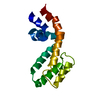


 PDBj
PDBj


 Assembly

 Sample preparation
 Processing
 X-PLOR