 Movie
Movie Controller

[English] 日本語
 Yorodumi
- PDB-1ex6: CRYSTAL STRUCTURE OF UNLIGANDED FORM OF GUANYLATE KINASE FROM YEAST -
+ Open data

- Basic information
| Entry | Database: PDB / ID: 1ex6 | ||||||
|---|---|---|---|---|---|---|---|
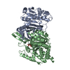
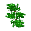
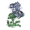
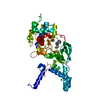
| Title | CRYSTAL STRUCTURE OF UNLIGANDED FORM OF GUANYLATE KINASE FROM YEAST | ||||||
 Components Components | GUANYLATE KINASE | ||||||
 Keywords Keywords | TRANSFERASE / GUANYLATE KINASE / SUBSTRATE-INDUCED FIT / DOMAIN MOVEMENT / ATP / GMP / SUBSTRATE SPECIFICITY | ||||||
| Function / homology |  Function and homology information Function and homology informationAzathioprine ADME / GDP biosynthetic process / guanylate kinase / purine nucleotide metabolic process / Interconversion of nucleotide di- and triphosphates / GMP kinase activity / ATP binding / nucleus / cytoplasm / cytosol Similarity search - Function | ||||||
| Biological species |  | ||||||
| Method |  X-RAY DIFFRACTION / SYNCHROTRON / HGMAD / Resolution: 2.3 Å X-RAY DIFFRACTION / SYNCHROTRON / HGMAD / Resolution: 2.3 Å | ||||||
 Authors Authors | Blaszczyk, J. / Ji, X. | ||||||
 Citation Citation | Journal: J.Mol.Biol. / Year: 2001 Title: Crystal structure of unligated guanylate kinase from yeast reveals GMP-induced conformational changes. Authors: Blaszczyk, J. / Li, Y. / Yan, H. / Ji, X. | ||||||
| History |
|
- Structure visualization
| Structure viewer | Molecule: MolmilJmol/JSmol |
|---|
- Downloads & links
-Download
| PDBx/mmCIF format | 1ex6.cif.gz | 93 KB | Display | PDBx/mmCIF format |
|---|---|---|---|---|
| PDB format | pdb1ex6.ent.gz | 70.4 KB | Display | PDB format |
| PDBx/mmJSON format | 1ex6.json.gz | Tree view | PDBx/mmJSON format | |
| Others |  Other downloads Other downloads |
-Validation report
| Summary document | 1ex6_validation.pdf.gz | 426.3 KB | Display | wwPDB validaton report |
|---|---|---|---|---|
| Full document | 1ex6_full_validation.pdf.gz | 442.5 KB | Display | |
| Data in XML | 1ex6_validation.xml.gz | 22 KB | Display | |
| Data in CIF | 1ex6_validation.cif.gz | 31.9 KB | Display | |
| Arichive directory | https://data.pdbj.org/pub/pdb/validation_reports/ex/1ex6ftp://data.pdbj.org/pub/pdb/validation_reports/ex/1ex6 | HTTPS FTP |
-Related structure data


-Links
 PDBj
PDBj- Assembly
| Deposited unit | 
| ||||||||
|---|---|---|---|---|---|---|---|---|---|
| 1 |
| ||||||||
| Unit cell |
|
-Components
| #1: Protein | Mass: 20533.152 Da / Num. of mol.: 2 Source method: isolated from a genetically manipulated source Source: (gene. exp.) Plasmid: PET17B / Species (production host): Escherichia coli / Production host:  #2: Water | ChemComp-HOH / |  Mass: 18.015 Da / Num. of mol.: 424 / Source method: isolated from a natural source / Formula: H2O Mass: 18.015 Da / Num. of mol.: 424 / Source method: isolated from a natural source / Formula: H2O |
|---|
-Experimental details
-Experiment
| Experiment | Method: X-RAY DIFFRACTION / Number of used crystals: 1 |
|---|
- Sample preparation
| Crystal | Density Matthews: 2.1 Å3/Da / Density % sol: 39.9 % | ||||||||||||||||||||||||||||||
|---|---|---|---|---|---|---|---|---|---|---|---|---|---|---|---|---|---|---|---|---|---|---|---|---|---|---|---|---|---|---|---|
| Crystal grow | Temperature: 292 K / Method: vapor diffusion, hanging drop / pH: 8.5 Details: PEG4000, Tris-HCl, acetate, pH 8.5, VAPOR DIFFUSION, HANGING DROP, temperature 292K | ||||||||||||||||||||||||||||||
| Crystal grow | *PLUS Temperature: 100 K / pH: 7 Details: drop consists of equal amounts of protein and reservoir solutions | ||||||||||||||||||||||||||||||
| Components of the solutions | *PLUS
|
-Data collection
| Diffraction | Mean temperature: 100 K |
|---|---|
| Diffraction source | Source: SYNCHROTRON / Site: NSLS  / Beamline: X9B / Wavelength: 0.92 / Beamline: X9B / Wavelength: 0.92 |
| Detector | Type: ADSC QUANTUM 4 / Detector: CCD / Date: Mar 24, 1998 / Details: MIRROR |
| Radiation | Monochromator: SILICON 111 / Protocol: MAD / Monochromatic (M) / Laue (L): M / Scattering type: x-ray |
| Radiation wavelength | Wavelength: 0.92 Å / Relative weight: 1 |
| Reflection | Resolution: 2.3→20 Å / Num. all: 15759 / Num. obs: 15759 / % possible obs: 99.7 % / Observed criterion σ(F): 0 / Observed criterion σ(I): 0 / Redundancy: 2.37 % / Biso Wilson estimate: 38.9 Å2 / Rmerge(I) obs: 0.08 / Net I/σ(I): 9.9917 |
| Reflection shell | Resolution: 2.3→2.34 Å / Redundancy: 2.13 % / Rmerge(I) obs: 0.543 / Mean I/σ(I) obs: 1.5845 / Num. unique all: 776 / % possible all: 97.9 |
| Reflection | *PLUS Num. measured all: 37285 |
| Reflection shell | *PLUS % possible obs: 97.9 % |
- Processing
| Software |
| |||||||||||||||||||||||||||||||||
|---|---|---|---|---|---|---|---|---|---|---|---|---|---|---|---|---|---|---|---|---|---|---|---|---|---|---|---|---|---|---|---|---|---|---|
| Refinement | Method to determine structure: HGMAD / Resolution: 2.3→20 Å / Num. parameters: 11965 / Num. restraintsaints: 11875 / Cross valid method: FREE R / σ(F): 4 / σ(I): 2 / Stereochemistry target values: ENGH AND HUBER Details: Least-squares refinement using the Konnert-Hendrickson conjugate-gradient algorithm
| |||||||||||||||||||||||||||||||||
| Solvent computation | Solvent model: MOEWS & KRETSINGER, J.MOL.BIOL.91(1975)201-228 | |||||||||||||||||||||||||||||||||
| Refine analyze | Occupancy sum hydrogen: 0 / Occupancy sum non hydrogen: 3306 | |||||||||||||||||||||||||||||||||
| Refinement step | Cycle: LAST / Resolution: 2.3→20 Å
| |||||||||||||||||||||||||||||||||
| Refine LS restraints |
| |||||||||||||||||||||||||||||||||
| Software | *PLUS Name: SHELXL-97 / Classification: refinement | |||||||||||||||||||||||||||||||||
| Refinement | *PLUS Rfactor all: 0.214 / Rfactor obs: 0.164 | |||||||||||||||||||||||||||||||||
| Solvent computation | *PLUS | |||||||||||||||||||||||||||||||||
| Displacement parameters | *PLUS |
