 Movie
Movie Controller


+ Open data

- Basic information
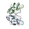
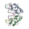
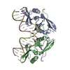
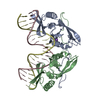
| Entry | Database: PDB / ID: 1evx | ||||||
|---|---|---|---|---|---|---|---|
| Title | APO CRYSTAL STRUCTURE OF THE HOMING ENDONUCLEASE, I-PPOI | ||||||
 Components Components | INTRON-ENCODED HOMING ENDONUCLEASE I-PPOI | ||||||
 Keywords Keywords | HYDROLASE / DNA binding B-sheets / C-terminal exchanged dimer interface | ||||||
| Function / homology |  Function and homology information Function and homology informationintron homing / endonuclease activity / Hydrolases; Acting on ester bonds / hydrolase activity Similarity search - Function | ||||||
| Biological species |  Physarum polycephalum (eukaryote) Physarum polycephalum (eukaryote) | ||||||
| Method |  X-RAY DIFFRACTION / SYNCHROTRON / Resolution: 2 Å X-RAY DIFFRACTION / SYNCHROTRON / Resolution: 2 Å | ||||||
 Authors Authors | Galburt, E.A. / Jurica, M.S. / Chevalier, B.S. / Erho, D. / Stoddard, B.L. | ||||||
 Citation Citation | Journal: J.Mol.Biol. / Year: 2000 Title: Conformational changes and cleavage by the homing endonuclease I-PpoI: a critical role for a leucine residue in the active site. Authors: Galburt, E.A. / Chadsey, M.S. / Jurica, M.S. / Chevalier, B.S. / Erho, D. / Tang, W. / Monnat Jr., R.J. / Stoddard, B.L. | ||||||
| History |
|
- Structure visualization
| Structure viewer | Molecule: MolmilJmol/JSmol |
|---|
- Downloads & links
-Download
| PDBx/mmCIF format | 1evx.cif.gz | 82 KB | Display | PDBx/mmCIF format |
|---|---|---|---|---|
| PDB format | pdb1evx.ent.gz | 61.1 KB | Display | PDB format |
| PDBx/mmJSON format | 1evx.json.gz | Tree view | PDBx/mmJSON format | |
| Others |  Other downloads Other downloads |
-Validation report
| Arichive directory | https://data.pdbj.org/pub/pdb/validation_reports/ev/1evxftp://data.pdbj.org/pub/pdb/validation_reports/ev/1evx | HTTPS FTP |
|---|
-Related structure data


-Links
 PDBj
PDBj- Assembly
| Deposited unit | 
| ||||||||
|---|---|---|---|---|---|---|---|---|---|
| 1 |
| ||||||||
| Unit cell |
| ||||||||
| Components on special symmetry positions |
| ||||||||
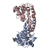
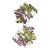
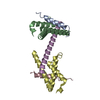
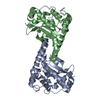
| Details | The biological assembly is the dimer of chain A and chain B. |
-Components
| #1: Protein | Mass: 17680.047 Da / Num. of mol.: 2 Source method: isolated from a genetically manipulated source Source: (gene. exp.) Physarum polycephalum (eukaryote) / Production host:  #2: Chemical | ChemComp-ZN /   Mass: 65.409 Da / Num. of mol.: 4 / Source method: obtained synthetically / Formula: Zn Mass: 65.409 Da / Num. of mol.: 4 / Source method: obtained synthetically / Formula: Zn#3: Chemical | ChemComp-SO4 /   Mass: 96.063 Da / Num. of mol.: 4 / Source method: obtained synthetically / Formula: SO4 Mass: 96.063 Da / Num. of mol.: 4 / Source method: obtained synthetically / Formula: SO4#4: Water | ChemComp-HOH / |  Mass: 18.015 Da / Num. of mol.: 310 / Source method: isolated from a natural source / Formula: H2O Mass: 18.015 Da / Num. of mol.: 310 / Source method: isolated from a natural source / Formula: H2O |
|---|
-Experimental details
-Experiment
| Experiment | Method: X-RAY DIFFRACTION / Number of used crystals: 1 |
|---|
- Sample preparation
| Crystal | Density Matthews: 2.75 Å3/Da / Density % sol: 55.2 % | |||||||||||||||||||||||||
|---|---|---|---|---|---|---|---|---|---|---|---|---|---|---|---|---|---|---|---|---|---|---|---|---|---|---|
| Crystal grow | Temperature: 277 K / Method: vapor diffusion, hanging drop / pH: 6.5 Details: ammonium sulfate, PEG 8K, sodium cacodylate pH 6.5, VAPOR DIFFUSION, HANGING DROP, temperature 4K | |||||||||||||||||||||||||
| Crystal grow | *PLUS | |||||||||||||||||||||||||
| Components of the solutions | *PLUS
|
-Data collection
| Diffraction | Mean temperature: 77 K |
|---|---|
| Diffraction source | Source: SYNCHROTRON / Site: ALS  / Beamline: 5.0.2 / Wavelength: 0.98 / Beamline: 5.0.2 / Wavelength: 0.98 |
| Detector | Detector: CCD |
| Radiation | Protocol: SINGLE WAVELENGTH / Monochromatic (M) / Laue (L): M / Scattering type: x-ray |
| Radiation wavelength | Wavelength: 0.98 Å / Relative weight: 1 |
| Reflection | Resolution: 2→50 Å / Num. all: 102030 / Num. obs: 25509 / % possible obs: 90.8 % / Observed criterion σ(F): 0 / Observed criterion σ(I): 0 / Redundancy: 4 % / Rmerge(I) obs: 0.052 / Net I/σ(I): 12.2 |
| Reflection shell | Resolution: 2→2.03 Å / Rmerge(I) obs: 0.204 / % possible all: 74.8 |
| Reflection shell | *PLUS % possible obs: 74.8 % / Mean I/σ(I) obs: 7 |
- Processing
| Software |
| ||||||||||||||||||||
|---|---|---|---|---|---|---|---|---|---|---|---|---|---|---|---|---|---|---|---|---|---|
| Refinement | Resolution: 2→50 Å / σ(F): 0 / σ(I): 0
| ||||||||||||||||||||
| Refinement step | Cycle: LAST / Resolution: 2→50 Å
| ||||||||||||||||||||
| Refine LS restraints |
| ||||||||||||||||||||
| Software | *PLUS Name: X-PLOR / Version: 3.851 / Classification: refinement | ||||||||||||||||||||
| Refinement | *PLUS | ||||||||||||||||||||
| Solvent computation | *PLUS | ||||||||||||||||||||
| Displacement parameters | *PLUS Biso mean: 26.9 Å2 | ||||||||||||||||||||
| Refine LS restraints | *PLUS
|
