 Movie
Movie Controller


+ Open data

- Basic information
| Entry | Database: PDB / ID: 1evh | ||||||
|---|---|---|---|---|---|---|---|
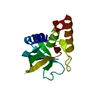
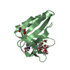
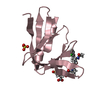
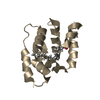
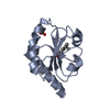
| Title | EVH1 DOMAIN FROM MURINE ENABLED IN COMPLEX WITH ACTA PEPTIDE | ||||||
 Components Components |
| ||||||
 Keywords Keywords | CONTRACTILE PROTEIN / MOLECULAR RECOGNITION / ACTIN DYNAMICS | ||||||
| Function / homology |  Function and homology information Function and homology informationSignaling by ROBO receptors / postsynaptic cytoskeleton organization / actin polymerization-dependent cell motility / profilin binding / actin polymerization or depolymerization / WW domain binding / cellular response to leukemia inhibitory factor / stress fiber / axon guidance / neural tube closure ...Signaling by ROBO receptors / postsynaptic cytoskeleton organization / actin polymerization-dependent cell motility / profilin binding / actin polymerization or depolymerization / WW domain binding / cellular response to leukemia inhibitory factor / stress fiber / axon guidance / neural tube closure / actin filament organization / filopodium / SH3 domain binding / GABA-ergic synapse / lamellipodium / actin cytoskeleton / actin cytoskeleton organization / actin binding / postsynapse / focal adhesion / plasma membrane / cytosol Similarity search - Function | ||||||
| Biological species |  | ||||||
| Method |  X-RAY DIFFRACTION / SYNCHROTRON / MAD / Resolution: 1.8 Å X-RAY DIFFRACTION / SYNCHROTRON / MAD / Resolution: 1.8 Å | ||||||
 Authors Authors | Prehoda, K.E. / Lee, D.J. / Lim, W.A. | ||||||
 Citation Citation | Journal: Cell(Cambridge,Mass.) / Year: 1999 Title: Structure of the enabled/VASP homology 1 domain-peptide complex: a key component in the spatial control of actin assembly. Authors: Prehoda, K.E. / Lee, D.J. / Lim, W.A. | ||||||
| History |
|
- Structure visualization
| Structure viewer | Molecule: MolmilJmol/JSmol |
|---|
- Downloads & links
-Download
| PDBx/mmCIF format | 1evh.cif.gz | 35.7 KB | Display | PDBx/mmCIF format |
|---|---|---|---|---|
| PDB format | pdb1evh.ent.gz | 23.6 KB | Display | PDB format |
| PDBx/mmJSON format | 1evh.json.gz | Tree view | PDBx/mmJSON format | |
| Others |  Other downloads Other downloads |
-Validation report
| Arichive directory | https://data.pdbj.org/pub/pdb/validation_reports/ev/1evhftp://data.pdbj.org/pub/pdb/validation_reports/ev/1evh | HTTPS FTP |
|---|
-Related structure data
| Similar structure data |
|---|

-Links
 PDBj
PDBj- Assembly
| Deposited unit | 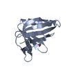
| ||||||||||
|---|---|---|---|---|---|---|---|---|---|---|---|
| 1 |
| ||||||||||
| Unit cell |
|
-Components
| #1: Protein | Mass: 12598.247 Da / Num. of mol.: 1 / Fragment: MENA 1-112 Source method: isolated from a genetically manipulated source Source: (gene. exp.)  |
|---|---|
| #2: Protein/peptide | Mass: 680.790 Da / Num. of mol.: 1 / Fragment: PROLINE RICH REPEAT / Source method: obtained synthetically / Details: sequence from LISTERIA MONOCYTOGENES |
| #3: Water | ChemComp-HOH /  Mass: 18.015 Da / Num. of mol.: 27 / Source method: isolated from a natural source / Formula: H2O Mass: 18.015 Da / Num. of mol.: 27 / Source method: isolated from a natural source / Formula: H2O |
| Has protein modification | Y |
-Experimental details
-Experiment
| Experiment | Method: X-RAY DIFFRACTION / Number of used crystals: 1 |
|---|
- Sample preparation
| Crystal | Density Matthews: 1.87 Å3/Da / Density % sol: 34.1 % | |||||||||||||||||||||||||
|---|---|---|---|---|---|---|---|---|---|---|---|---|---|---|---|---|---|---|---|---|---|---|---|---|---|---|
| Crystal grow | pH: 7.5 Details: 0.1 M NA HEPES, 70% SATURATED AMMONIUM PHOSPHATE, pH 7.5 | |||||||||||||||||||||||||
| Crystal grow | *PLUS Temperature: 20 ℃ / Method: vapor diffusion, sitting drop | |||||||||||||||||||||||||
| Components of the solutions | *PLUS
|
-Data collection
| Diffraction | Mean temperature: 200 K | |||||||||
|---|---|---|---|---|---|---|---|---|---|---|
| Diffraction source | Source: SYNCHROTRON / Site: ALS  / Beamline: 5.0.2 / Wavelength: 0.96859, 0.99984 / Beamline: 5.0.2 / Wavelength: 0.96859, 0.99984 | |||||||||
| Detector | Type: ADSC / Detector: CCD / Date: Nov 15, 1998 | |||||||||
| Radiation | Monochromator: SI FILTER / Protocol: MAD / Monochromatic (M) / Laue (L): M / Scattering type: x-ray | |||||||||
| Radiation wavelength |
| |||||||||
| Reflection | Resolution: 1.8→30 Å / Num. obs: 17747 / % possible obs: 99.8 % / Observed criterion σ(I): 0 / Redundancy: 6 % / Biso Wilson estimate: 14.9 Å2 / Rsym value: 9 / Net I/σ(I): 11 | |||||||||
| Reflection shell | Resolution: 1.8→1.83 Å / Rsym value: 22 / % possible all: 97.6 | |||||||||
| Reflection | *PLUS % possible obs: 99.1 % / Num. measured all: 88290 / Rmerge(I) obs: 0.09 | |||||||||
| Reflection shell | *PLUS % possible obs: 91.4 % / Rmerge(I) obs: 0.358 |
- Processing
| Software |
| ||||||||||||||||||||||||||||||||||||||||||||||||||||||||||||
|---|---|---|---|---|---|---|---|---|---|---|---|---|---|---|---|---|---|---|---|---|---|---|---|---|---|---|---|---|---|---|---|---|---|---|---|---|---|---|---|---|---|---|---|---|---|---|---|---|---|---|---|---|---|---|---|---|---|---|---|---|---|
| Refinement | Method to determine structure: MAD / Resolution: 1.8→30 Å / Rfactor Rfree error: 0.009 / Cross valid method: THROUGHOUT / σ(F): 0
| ||||||||||||||||||||||||||||||||||||||||||||||||||||||||||||
| Solvent computation | Solvent model: FLAT MODEL / Bsol: 45.47 Å2 / ksol: 0.38 e/Å3 | ||||||||||||||||||||||||||||||||||||||||||||||||||||||||||||
| Displacement parameters | Biso mean: 25.7 Å2
| ||||||||||||||||||||||||||||||||||||||||||||||||||||||||||||
| Refine analyze |
| ||||||||||||||||||||||||||||||||||||||||||||||||||||||||||||
| Refinement step | Cycle: LAST / Resolution: 1.8→30 Å
| ||||||||||||||||||||||||||||||||||||||||||||||||||||||||||||
| Refine LS restraints |
| ||||||||||||||||||||||||||||||||||||||||||||||||||||||||||||
| LS refinement shell | Resolution: 1.8→1.91 Å / Rfactor Rfree error: 0.022 / Total num. of bins used: 6
| ||||||||||||||||||||||||||||||||||||||||||||||||||||||||||||
| Xplor file |
| ||||||||||||||||||||||||||||||||||||||||||||||||||||||||||||
| Software | *PLUS Name: CNS / Version: 0.4 / Classification: refinement | ||||||||||||||||||||||||||||||||||||||||||||||||||||||||||||
| Refinement | *PLUS Highest resolution: 1.8 Å / Lowest resolution: 30 Å / σ(F): 0 / % reflection Rfree: 10.2 % / Rfactor obs: 0.229 / Rfactor Rwork: 0.23 | ||||||||||||||||||||||||||||||||||||||||||||||||||||||||||||
| Solvent computation | *PLUS | ||||||||||||||||||||||||||||||||||||||||||||||||||||||||||||
| Displacement parameters | *PLUS Biso mean: 25.7 Å2 | ||||||||||||||||||||||||||||||||||||||||||||||||||||||||||||
| Refine LS restraints | *PLUS
| ||||||||||||||||||||||||||||||||||||||||||||||||||||||||||||
| LS refinement shell | *PLUS Highest resolution: 1.8 Å / Rfactor Rfree: 0.286 / % reflection Rfree: 11.3 % / Rfactor Rwork: 0.27 |
