 Movie
Movie Controller


+ Open data

- Basic information
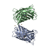
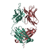
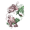
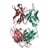
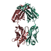
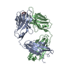
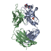
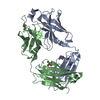
| Entry | Database: PDB / ID: 1es8 | ||||||
|---|---|---|---|---|---|---|---|
| Title | Crystal structure of free BglII | ||||||
 Components Components | RESTRICTION ENDONUCLEASE BGLII | ||||||
 Keywords Keywords | HYDROLASE / RESTRICTION ENDONUCLEASE / RESTRICTION ENZYME / uncomplexed | ||||||
| Function / homology |  Function and homology information Function and homology informationtype II site-specific deoxyribonuclease / type II site-specific deoxyribonuclease activity / DNA restriction-modification system / magnesium ion binding / DNA binding Similarity search - Function | ||||||
| Biological species |  | ||||||
| Method |  X-RAY DIFFRACTION / SYNCHROTRON / MAD / Resolution: 2.3 Å X-RAY DIFFRACTION / SYNCHROTRON / MAD / Resolution: 2.3 Å | ||||||
 Authors Authors | Lukacs, C.M. / Aggarwal, A.K. | ||||||
 Citation Citation | Journal: Nat.Struct.Biol. / Year: 2001 Title: Structure of free BglII reveals an unprecedented scissor-like motion for opening an endonuclease. Authors: Lukacs, C.M. / Kucera, R. / Schildkraut, I. / Aggarwal, A.K. | ||||||
| History |
|
- Structure visualization
| Structure viewer | Molecule: MolmilJmol/JSmol |
|---|
- Downloads & links
-Download
| PDBx/mmCIF format | 1es8.cif.gz | 53.7 KB | Display | PDBx/mmCIF format |
|---|---|---|---|---|
| PDB format | pdb1es8.ent.gz | 39.3 KB | Display | PDB format |
| PDBx/mmJSON format | 1es8.json.gz | Tree view | PDBx/mmJSON format | |
| Others |  Other downloads Other downloads |
-Validation report
| Arichive directory | https://data.pdbj.org/pub/pdb/validation_reports/es/1es8ftp://data.pdbj.org/pub/pdb/validation_reports/es/1es8 | HTTPS FTP |
|---|
-Related structure data
| Related structure data | |
|---|---|
| Similar structure data |


-Links
 PDBj
PDBj


- Assembly
| Deposited unit | 
| ||||||||
|---|---|---|---|---|---|---|---|---|---|
| 1 | 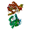
| ||||||||
| Unit cell |
|
-Components
| #1: Protein | Mass: 26031.756 Da / Num. of mol.: 1 Source method: isolated from a genetically manipulated source Source: (gene. exp.) References: UniProt: Q45488, type II site-specific deoxyribonuclease |
|---|---|
| #2: Chemical | ChemComp-ACY /   Mass: 60.052 Da / Num. of mol.: 1 / Source method: obtained synthetically / Formula: C2H4O2 Mass: 60.052 Da / Num. of mol.: 1 / Source method: obtained synthetically / Formula: C2H4O2 |
| #3: Water | ChemComp-HOH /  Mass: 18.015 Da / Num. of mol.: 81 / Source method: isolated from a natural source / Formula: H2O Mass: 18.015 Da / Num. of mol.: 81 / Source method: isolated from a natural source / Formula: H2O |
| Has protein modification | Y |
-Experimental details
-Experiment
| Experiment | Method: X-RAY DIFFRACTION / Number of used crystals: 1 |
|---|
- Sample preparation
| Crystal | Density Matthews: 2.31 Å3/Da / Density % sol: 46.75 % | |||||||||||||||||||||||||||||||||||||||||||||||||||||||||||||||
|---|---|---|---|---|---|---|---|---|---|---|---|---|---|---|---|---|---|---|---|---|---|---|---|---|---|---|---|---|---|---|---|---|---|---|---|---|---|---|---|---|---|---|---|---|---|---|---|---|---|---|---|---|---|---|---|---|---|---|---|---|---|---|---|---|
| Crystal grow | Temperature: 277 K / Method: vapor diffusion, hanging drop / pH: 6 Details: 30% isopropanol, 0.2 M sodium acetate, 0.1 M Bis-tris, pH 6.0, VAPOR DIFFUSION, HANGING DROP, temperature 277K | |||||||||||||||||||||||||||||||||||||||||||||||||||||||||||||||
| Crystal grow | *PLUS Temperature: 4 ℃ / pH: 7.4 Details: drop consists of equal amounts of protein and precipitant solutions | |||||||||||||||||||||||||||||||||||||||||||||||||||||||||||||||
| Components of the solutions | *PLUS
|
-Data collection
| Diffraction | Mean temperature: 100 K | ||||||||||||
|---|---|---|---|---|---|---|---|---|---|---|---|---|---|
| Diffraction source | Source: SYNCHROTRON / Site: NSLS  / Beamline: X25 / Wavelength: 0.9793, 0.9789, 0.9689 / Beamline: X25 / Wavelength: 0.9793, 0.9789, 0.9689 | ||||||||||||
| Detector | Type: BRANDEIS - B4 / Detector: CCD / Date: Mar 1, 2000 | ||||||||||||
| Radiation | Protocol: MAD / Monochromatic (M) / Laue (L): M / Scattering type: x-ray | ||||||||||||
| Radiation wavelength |
| ||||||||||||
| Reflection | Resolution: 2.3→40 Å / Num. all: 10979 / Num. obs: 10979 / % possible obs: 99.4 % / Observed criterion σ(I): -2 / Redundancy: 3.1 % / Biso Wilson estimate: 36.6 Å2 / Rmerge(I) obs: 0.063 / Net I/σ(I): 14.4 | ||||||||||||
| Reflection shell | Resolution: 2.3→2.38 Å / Redundancy: 2.9 % / Rmerge(I) obs: 0.152 / Num. unique all: 1083 / % possible all: 99.1 | ||||||||||||
| Reflection | *PLUS Num. measured all: 33794 / Rmerge(I) obs: 0.057 | ||||||||||||
| Reflection shell | *PLUS % possible obs: 99.1 % / Rmerge(I) obs: 0.138 |
- Processing
| Software |
| |||||||||||||||||||||||||
|---|---|---|---|---|---|---|---|---|---|---|---|---|---|---|---|---|---|---|---|---|---|---|---|---|---|---|
| Refinement | Method to determine structure: MAD / Resolution: 2.3→20 Å / Cross valid method: THROUGHOUT / σ(F): 0 / σ(I): 0 / Stereochemistry target values: Engh & Huber
| |||||||||||||||||||||||||
| Refinement step | Cycle: LAST / Resolution: 2.3→20 Å
| |||||||||||||||||||||||||
| Refine LS restraints |
| |||||||||||||||||||||||||
| Software | *PLUS Name: CNS / Classification: refinement | |||||||||||||||||||||||||
| Refinement | *PLUS Num. reflection all: 10418 | |||||||||||||||||||||||||
| Solvent computation | *PLUS | |||||||||||||||||||||||||
| Displacement parameters | *PLUS Biso mean: 37.1 Å2 |
