 Movie
Movie Controller

[English] 日本語
 Yorodumi
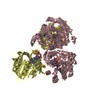
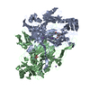
- PDB-1ep3: CRYSTAL STRUCTURE OF LACTOCOCCUS LACTIS DIHYDROOROTATE DEHYDROGEN... -
+ Open data

- Basic information
| Entry | Database: PDB / ID: 1ep3 | ||||||
|---|---|---|---|---|---|---|---|
| Title | CRYSTAL STRUCTURE OF LACTOCOCCUS LACTIS DIHYDROOROTATE DEHYDROGENASE B. DATA COLLECTED UNDER CRYOGENIC CONDITIONS. | ||||||
 Components Components | (DIHYDROOROTATE DEHYDROGENASE B ...) x 2 | ||||||
 Keywords Keywords | OXIDOREDUCTASE / Heterotetramer / ALPHA-BETA barrel / BETA sandwich / FAD domain / ALPHA/BETA NADP domain / FeS cluster binding domain | ||||||
| Function / homology |  Function and homology information Function and homology informationdihydroorotate dehydrogenase (NAD+) / dihydroorotate dehydrogenase (NAD+) activity / 'de novo' UMP biosynthetic process / 'de novo' pyrimidine nucleobase biosynthetic process / 2 iron, 2 sulfur cluster binding / flavin adenine dinucleotide binding / electron transfer activity / oxidoreductase activity / metal ion binding / cytoplasm Similarity search - Function | ||||||
| Biological species |  Lactococcus lactis (lactic acid bacteria) Lactococcus lactis (lactic acid bacteria) | ||||||
| Method |  X-RAY DIFFRACTION / SYNCHROTRON / MOLECULAR REPLACEMENT / Resolution: 2.1 Å X-RAY DIFFRACTION / SYNCHROTRON / MOLECULAR REPLACEMENT / Resolution: 2.1 Å | ||||||
 Authors Authors | Rowland, P. / Norager, S. / Jensen, K.F. / Larsen, S. | ||||||
 Citation Citation | Journal: Structure Fold.Des. / Year: 2000 Title: Structure of dihydroorotate dehydrogenase B: electron transfer between two flavin groups bridged by an iron-sulphur cluster. Authors: Rowland, P. / Norager, S. / Jensen, K.F. / Larsen, S. #1: Journal: Acta Crystallogr.,Sect.D / Year: 1997Title: Crystallization and Preliminary X-ray Diffraction Analysis of the Heterotetrameric Dihydroorotate Dehydrogenase B of Lactococcus lactis, a Flavoprotein Enzyme System Consisting of two PyrDB ...Title: Crystallization and Preliminary X-ray Diffraction Analysis of the Heterotetrameric Dihydroorotate Dehydrogenase B of Lactococcus lactis, a Flavoprotein Enzyme System Consisting of two PyrDB Subunits and Two Iron-Sulfur Cluster Containing PyrK Subunits Authors: Rowland, P. / Nielsen, F.S. / Jensen, K.F. / Larsen, S. #2: Journal: J.Biol.Chem. / Year: 1996Title: The B Form of Dihydroorotate Dehydrogenase from Lactococcus lactis Consists of two Different Subunits, Encoded by the pyrDb and PyrK Genes, and Contains FMN, FAD and [FeS] Redox Centers Authors: Nielsen, F.S. / Andersen, P.S. / Jensen, K.F. #3: Journal: Flavins and Flavoproteins / Year: 1999Title: Roles of three prosthetic groups in tetrameric Dihydroorotate dehydrogenase B from Lactococcus lactis Authors: Jensen, K.F. / Bjornberg, O. / Nielsen, F.S. / Ottosen, M. / Sorensen, P.G. / Rowland, P. / Norager, S. / Larsen, S. | ||||||
| History |
|
- Structure visualization
| Structure viewer | Molecule: MolmilJmol/JSmol |
|---|
- Downloads & links
-Download
| PDBx/mmCIF format | 1ep3.cif.gz | 137.2 KB | Display | PDBx/mmCIF format |
|---|---|---|---|---|
| PDB format | pdb1ep3.ent.gz | 104.2 KB | Display | PDB format |
| PDBx/mmJSON format | 1ep3.json.gz | Tree view | PDBx/mmJSON format | |
| Others |  Other downloads Other downloads |
-Validation report
| Arichive directory | https://data.pdbj.org/pub/pdb/validation_reports/ep/1ep3ftp://data.pdbj.org/pub/pdb/validation_reports/ep/1ep3 | HTTPS FTP |
|---|
-Related structure data
| Related structure data | 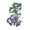 1ep1SC  1ep2C S: Starting model for refinement C: citing same article ( |
|---|---|
| Similar structure data |


-Links
 PDBj
PDBj


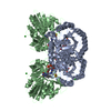
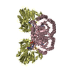
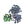
- Assembly
| Deposited unit | 
| ||||||||
|---|---|---|---|---|---|---|---|---|---|
| 1 | 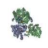
| ||||||||
| Unit cell |
| ||||||||
| Components on special symmetry positions |
|
-Components
-DIHYDROOROTATE DEHYDROGENASE B ... , 2 types, 2 molecules AB
| #1: Protein | Mass: 32977.289 Da / Num. of mol.: 1 Source method: isolated from a genetically manipulated source Source: (gene. exp.) Lactococcus lactis (lactic acid bacteria)Production host: |
|---|---|
| #2: Protein | Mass: 28690.295 Da / Num. of mol.: 1 Source method: isolated from a genetically manipulated source Source: (gene. exp.) Lactococcus lactis (lactic acid bacteria)Production host: |
-Non-polymers , 4 types, 469 molecules 


| #3: Chemical | ChemComp-FMN /  Mass: 456.344 Da / Num. of mol.: 1 / Source method: obtained synthetically / Formula: C17H21N4O9P Mass: 456.344 Da / Num. of mol.: 1 / Source method: obtained synthetically / Formula: C17H21N4O9P |
|---|---|
| #4: Chemical | ChemComp-FAD /  Mass: 785.550 Da / Num. of mol.: 1 / Source method: obtained synthetically / Formula: C27H33N9O15P2 / Comment: FAD*YM Mass: 785.550 Da / Num. of mol.: 1 / Source method: obtained synthetically / Formula: C27H33N9O15P2 / Comment: FAD*YM |
| #5: Chemical | ChemComp-FES /  Mass: 175.820 Da / Num. of mol.: 1 / Source method: obtained synthetically / Formula: Fe2S2 Mass: 175.820 Da / Num. of mol.: 1 / Source method: obtained synthetically / Formula: Fe2S2 |
| #6: Water | ChemComp-HOH / Mass: 18.015 Da / Num. of mol.: 466 / Source method: isolated from a natural source / Formula: H2O |
-Experimental details
-Experiment
| Experiment | Method: X-RAY DIFFRACTION / Number of used crystals: 1 |
|---|
- Sample preparation
| Crystal | Density Matthews: 2.5 Å3/Da / Density % sol: 50.82 % | ||||||||||||||||||||||||||||||
|---|---|---|---|---|---|---|---|---|---|---|---|---|---|---|---|---|---|---|---|---|---|---|---|---|---|---|---|---|---|---|---|
| Crystal grow | Temperature: 298 K / Method: vapor diffusion, hanging drop / pH: 4.5 Details: 2.1 - 2.4 M Ammonium sulfate, 0.1 M Na-acetate pH 4 to 5, pH 4.5, VAPOR DIFFUSION, HANGING DROP, temperature 298K | ||||||||||||||||||||||||||||||
| Crystal grow | *PLUS pH: 6 Details: Rowland, P., (1997) Acta Crystallogr., Sect.D, 53, 802. | ||||||||||||||||||||||||||||||
| Components of the solutions | *PLUS
|
-Data collection
| Diffraction | Mean temperature: 120 K |
|---|---|
| Diffraction source | Source: SYNCHROTRON / Site: EMBL/DESY, HAMBURG  / Beamline: BW7B / Wavelength: 0.8469 / Beamline: BW7B / Wavelength: 0.8469 |
| Detector | Type: MARRESEARCH / Detector: IMAGE PLATE / Date: Apr 26, 1999 |
| Radiation | Protocol: SINGLE WAVELENGTH / Monochromatic (M) / Laue (L): M / Scattering type: x-ray |
| Radiation wavelength | Wavelength: 0.8469 Å / Relative weight: 1 |
| Reflection | Resolution: 2.1→50 Å / Num. all: 270295 / Num. obs: 35821 / % possible obs: 98.7 % / Observed criterion σ(F): 0 / Observed criterion σ(I): 0 / Redundancy: 7.5 % / Biso Wilson estimate: 17.5 Å2 / Rmerge(I) obs: 0.073 / Net I/σ(I): 16.62 |
| Reflection shell | Resolution: 2.1→2.14 Å / Rmerge(I) obs: 0.327 / Num. unique all: 1554 / % possible all: 87.5 |
| Reflection | *PLUS Num. measured all: 270295 |
- Processing
| Software |
| |||||||||||||||||||||||||
|---|---|---|---|---|---|---|---|---|---|---|---|---|---|---|---|---|---|---|---|---|---|---|---|---|---|---|
| Refinement | Method to determine structure: MOLECULAR REPLACEMENT Starting model: PDB ID 1EP1 Resolution: 2.1→20 Å / Rfactor Rfree error: 0.004 / Cross valid method: THROUGHOUT / σ(F): 0 / σ(I): 0 / Stereochemistry target values: Engh & Huber
| |||||||||||||||||||||||||
| Solvent computation | Solvent model: FLAT MODEL / Bsol: 79.1266 Å2 / ksol: 0.374949 e/Å3 | |||||||||||||||||||||||||
| Displacement parameters | Biso mean: 32.5 Å2 | |||||||||||||||||||||||||
| Refine analyze |
| |||||||||||||||||||||||||
| Refinement step | Cycle: LAST / Resolution: 2.1→20 Å
| |||||||||||||||||||||||||
| Refine LS restraints |
| |||||||||||||||||||||||||
| LS refinement shell | Resolution: 2.1→2.23 Å / Rfactor Rfree error: 0.012 / Total num. of bins used: 6
| |||||||||||||||||||||||||
| Xplor file | Serial no: 1 / Param file: protein_rep.param / Topol file: protein.top | |||||||||||||||||||||||||
| Software | *PLUS Name: CNS / Version: 0.9 / Classification: refinement | |||||||||||||||||||||||||
| Refinement | *PLUS Highest resolution: 2.1 Å / Lowest resolution: 20 Å / σ(F): 0 / % reflection Rfree: 10 % / Rfactor obs: 0.195 | |||||||||||||||||||||||||
| Solvent computation | *PLUS | |||||||||||||||||||||||||
| Displacement parameters | *PLUS Biso mean: 32.5 Å2 | |||||||||||||||||||||||||
| Refine LS restraints | *PLUS
| |||||||||||||||||||||||||
| LS refinement shell | *PLUS Rfactor Rfree: 0.295 / % reflection Rfree: 10.3 % / Rfactor Rwork: 0.251 |
