 Movie
Movie Controller


+ Open data

- Basic information
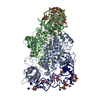
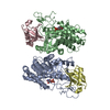
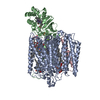
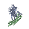
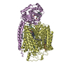
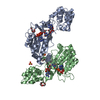
| Entry | Database: PDB / ID: 1eov | ||||||
|---|---|---|---|---|---|---|---|
| Title | FREE ASPARTYL-TRNA SYNTHETASE (ASPRS) (E.C. 6.1.1.12) FROM YEAST | ||||||
 Components Components | ASPARTYL-TRNA SYNTHETASE | ||||||
 Keywords Keywords | LIGASE / aminoacyl tRNA synthetase / tRNA ligase / apo-enzyme / OB-fold | ||||||
| Function / homology |  Function and homology information Function and homology informationaspartate-tRNA ligase / aspartate-tRNA ligase activity / aspartyl-tRNA aminoacylation / aminoacyl-tRNA synthetase multienzyme complex / sequence-specific mRNA binding / RNA binding / ATP binding / nucleus / cytoplasm / cytosol Similarity search - Function | ||||||
| Biological species |  | ||||||
| Method |  X-RAY DIFFRACTION / SYNCHROTRON / molecular replacement / Resolution: 2.3 Å X-RAY DIFFRACTION / SYNCHROTRON / molecular replacement / Resolution: 2.3 Å | ||||||
 Authors Authors | Sauter, C. / Lorber, B. / Cavarelli, J. / Moras, D. / Giege, R. | ||||||
 Citation Citation | Journal: J.Mol.Biol. / Year: 2000 Title: The free yeast aspartyl-tRNA synthetase differs from the tRNA(Asp)-complexed enzyme by structural changes in the catalytic site, hinge region, and anticodon-binding domain. Authors: Sauter, C. / Lorber, B. / Cavarelli, J. / Moras, D. / Giege, R. #1: Journal: Acta Crystallogr.,Sect.D / Year: 1999Title: Crystallogenesis Studies on Yeast Aspartyl-tRNA Synthetase: Use of Phase Diagram to Improve Crystal Quality. Authors: Sauter, C. / Lorber, B. / Kern, D. / Cavarelli, J. / Moras, D. / Giege, R. #2: Journal: Embo J. / Year: 1994Title: The Active Site of Yeast Aspartyl-tRNA Synthetase: Structural and Functional Aspects of the Aminoacylation Reaction. Authors: Cavarelli, J. / Eriani, G. / Rees, B. / Ruff, M. / Boeglin, M. / Mitschler, A. / Martin, F. / Gangloff, J. / Thierry, J.-C. / Moras, D. #3: Journal: Nature / Year: 1993Title: Yeast tRNA(Asp) Recognition by its Cognate Class II Aminoacyl-tRNA Synthetase. Authors: Cavarelli, J. / Rees, B. / Ruff, M. / Thierry, J.-C. / Moras, D. #4: Journal: Science / Year: 1991Title: Class II Aminoacyl Transfer RNA Synthetases: Crystal Structure of Yeast Aspartyl-tRNA Synthetase Complexed with tRNA(Asp). Authors: Ruff, M. / Krishnaswamy, S. / Boeglin, M. / Poterszman, A. / Mitschler, A. / Podjarny, A. / Rees, B. / Thierry, J.-C. / Moras, D. | ||||||
| History |
|
- Structure visualization
| Structure viewer | Molecule: MolmilJmol/JSmol |
|---|
- Downloads & links
-Download
| PDBx/mmCIF format | 1eov.cif.gz | 115.6 KB | Display | PDBx/mmCIF format |
|---|---|---|---|---|
| PDB format | pdb1eov.ent.gz | 88.2 KB | Display | PDB format |
| PDBx/mmJSON format | 1eov.json.gz | Tree view | PDBx/mmJSON format | |
| Others |  Other downloads Other downloads |
-Validation report
| Arichive directory | https://data.pdbj.org/pub/pdb/validation_reports/eo/1eovftp://data.pdbj.org/pub/pdb/validation_reports/eo/1eov | HTTPS FTP |
|---|
-Related structure data
| Related structure data | 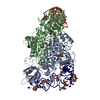 1aszS S: Starting model for refinement |
|---|---|
| Similar structure data |


-Links
 PDBj
PDBj

- Assembly
| Deposited unit | 
| ||||||||
|---|---|---|---|---|---|---|---|---|---|
| 1 | 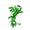
| ||||||||
| Unit cell |
| ||||||||
| Details | The biological assembly is a homodimer constructed from chain A and a symmetry partner generated by the two-fold. |
-Components
| #1: Protein | Mass: 55681.449 Da / Num. of mol.: 1 Fragment: ENGINEERED ASPRS MONOMER LACKING THE 70 N-TERMINAL AMINO ACID RESIDUES Source method: isolated from a genetically manipulated source Source: (gene. exp.) Gene: APS GENE / Plasmid: PTG908 / Production host:  |
|---|---|
| #2: Water | ChemComp-HOH /  Mass: 18.015 Da / Num. of mol.: 227 / Source method: isolated from a natural source / Formula: H2O Mass: 18.015 Da / Num. of mol.: 227 / Source method: isolated from a natural source / Formula: H2O |
-Experimental details
-Experiment
| Experiment | Method: X-RAY DIFFRACTION / Number of used crystals: 1 |
|---|
- Sample preparation
| Crystal | Density Matthews: 3.4 Å3/Da / Density % sol: 64 % | |||||||||||||||
|---|---|---|---|---|---|---|---|---|---|---|---|---|---|---|---|---|
| Crystal grow | Temperature: 278 K / Method: vapor diffusion, sitting drop / pH: 5.6 Details: 15 MG/ML PROTEIN, 1.9 M AMMONIUM SULFATE, pH 5.60, VAPOR DIFFUSION, SITTING DROP, temperature 278.0K | |||||||||||||||
| Crystal grow | *PLUS | |||||||||||||||
| Components of the solutions | *PLUS
|
-Data collection
| Diffraction | Mean temperature: 110 K |
|---|---|
| Diffraction source | Source: SYNCHROTRON / Site: ESRF  / Beamline: ID14-4 / Wavelength: 0.943 / Beamline: ID14-4 / Wavelength: 0.943 |
| Detector | Type: ADSC QUANTUM / Detector: CCD / Date: Feb 9, 1999 |
| Radiation | Protocol: SINGLE WAVELENGTH / Monochromatic (M) / Laue (L): M / Scattering type: x-ray |
| Radiation wavelength | Wavelength: 0.943 Å / Relative weight: 1 |
| Reflection | Resolution: 2.3→20 Å / Num. all: 183874 / Num. obs: 34124 / % possible obs: 98.2 % / Observed criterion σ(F): 0 / Observed criterion σ(I): -3 / Redundancy: 5.4 % / Biso Wilson estimate: 39.7 Å2 / Rsym value: 0.082 / Net I/σ(I): 12 |
| Reflection shell | Resolution: 2.3→2.35 Å / Redundancy: 5.5 % / Mean I/σ(I) obs: 4.6 / Rsym value: 0.277 / % possible all: 99.3 |
| Reflection | *PLUS Rmerge(I) obs: 0.082 |
| Reflection shell | *PLUS % possible obs: 99.3 % / Rmerge(I) obs: 0.277 |
- Processing
| Software |
| ||||||||||||||||||||||||||||||||||||||||||||||||||||||||||||
|---|---|---|---|---|---|---|---|---|---|---|---|---|---|---|---|---|---|---|---|---|---|---|---|---|---|---|---|---|---|---|---|---|---|---|---|---|---|---|---|---|---|---|---|---|---|---|---|---|---|---|---|---|---|---|---|---|---|---|---|---|---|
| Refinement | Method to determine structure: molecular replacement Starting model: 1ASZ Resolution: 2.3→20 Å / Rfactor Rfree error: 0.005 / Isotropic thermal model: OVERALL / Cross valid method: THROUGHOUT / σ(F): 0 / σ(I): 0 / Stereochemistry target values: ENGH & HUBER / Details: MAXIMUM-LIKELYHOOD TARGET USING AMPLITUDES.
| ||||||||||||||||||||||||||||||||||||||||||||||||||||||||||||
| Solvent computation | Solvent model: FLAT MODEL / Bsol: 37.29 Å2 / ksol: 0.331 e/Å3 | ||||||||||||||||||||||||||||||||||||||||||||||||||||||||||||
| Displacement parameters | Biso mean: 40.9 Å2
| ||||||||||||||||||||||||||||||||||||||||||||||||||||||||||||
| Refine analyze |
| ||||||||||||||||||||||||||||||||||||||||||||||||||||||||||||
| Refinement step | Cycle: LAST / Resolution: 2.3→20 Å
| ||||||||||||||||||||||||||||||||||||||||||||||||||||||||||||
| Refine LS restraints |
| ||||||||||||||||||||||||||||||||||||||||||||||||||||||||||||
| LS refinement shell | Resolution: 2.3→2.35 Å / Rfactor Rfree error: 0.024 / Total num. of bins used: 15
| ||||||||||||||||||||||||||||||||||||||||||||||||||||||||||||
| Xplor file |
| ||||||||||||||||||||||||||||||||||||||||||||||||||||||||||||
| Software | *PLUS Name: CNS / Version: 1 / Classification: refinement | ||||||||||||||||||||||||||||||||||||||||||||||||||||||||||||
| Refinement | *PLUS Num. reflection all: 30538 | ||||||||||||||||||||||||||||||||||||||||||||||||||||||||||||
| Solvent computation | *PLUS | ||||||||||||||||||||||||||||||||||||||||||||||||||||||||||||
| Displacement parameters | *PLUS | ||||||||||||||||||||||||||||||||||||||||||||||||||||||||||||
| Refine LS restraints | *PLUS
|
