 Movie
Movie Controller

[English] 日本語
 Yorodumi
- PDB-1enu: A new target for shigellosis: Rational design and crystallographi... -
+ Open data

- Basic information
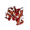
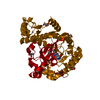
| Entry | Database: PDB / ID: 1enu | ||||||
|---|---|---|---|---|---|---|---|
| Title | A new target for shigellosis: Rational design and crystallographic studies of inhibitors of tRNA-guanine transglycosylase | ||||||
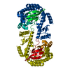
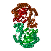
 Components Components | TRNA-GUANINE TRANSGLYCOSYLASE | ||||||
 Keywords Keywords | TRANSFERASE / TRNA-MODIFYING ENZYME / GLYCOSYLTRANSFERASE | ||||||
| Function / homology |  Function and homology information Function and homology informationtRNA-guanosine34 preQ1 transglycosylase / tRNA-guanosine(34) queuine transglycosylase activity / tRNA queuosine(34) biosynthetic process / metal ion binding / cytosol Similarity search - Function | ||||||
| Biological species |  Zymomonas mobilis (bacteria) Zymomonas mobilis (bacteria) | ||||||
| Method |  X-RAY DIFFRACTION / Resolution: 1.95 Å X-RAY DIFFRACTION / Resolution: 1.95 Å | ||||||
 Authors Authors | Gradler, U. / Gerber, H.D. / Goodenough-Lashua, D.M. / Garcia, G.A. / Ficner, R. / Reuter, K. / Stubbs, M.T. / Klebe, G. | ||||||
 Citation Citation | Journal: J.Mol.Biol. / Year: 2001 Title: A new target for shigellosis: rational design and crystallographic studies of inhibitors of tRNA-guanine transglycosylase. Authors: Gradler, U. / Gerber, H.D. / Goodenough-Lashua, D.M. / Garcia, G.A. / Ficner, R. / Reuter, K. / Stubbs, M.T. / Klebe, G. | ||||||
| History |
|
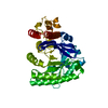
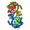
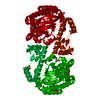
- Structure visualization
| Structure viewer | Molecule: MolmilJmol/JSmol |
|---|
- Downloads & links
-Download
| PDBx/mmCIF format | 1enu.cif.gz | 95.9 KB | Display | PDBx/mmCIF format |
|---|---|---|---|---|
| PDB format | pdb1enu.ent.gz | 71.3 KB | Display | PDB format |
| PDBx/mmJSON format | 1enu.json.gz | Tree view | PDBx/mmJSON format | |
| Others |  Other downloads Other downloads |
-Validation report
| Arichive directory | https://data.pdbj.org/pub/pdb/validation_reports/en/1enuftp://data.pdbj.org/pub/pdb/validation_reports/en/1enu | HTTPS FTP |
|---|
-Related structure data


-Links
 PDBj
PDBj- Assembly
| Deposited unit | 
| ||||||||
|---|---|---|---|---|---|---|---|---|---|
| 1 | 
| ||||||||
| Unit cell |
|
-Components
| #1: Protein | Mass: 42925.703 Da / Num. of mol.: 1 / Source method: isolated from a natural source / Source: (natural) Zymomonas mobilis (bacteria)References: UniProt: P28720, tRNA-guanosine34 preQ1 transglycosylase |
|---|---|
| #2: Chemical | ChemComp-ZN /   Mass: 65.409 Da / Num. of mol.: 1 / Source method: obtained synthetically / Formula: Zn Mass: 65.409 Da / Num. of mol.: 1 / Source method: obtained synthetically / Formula: Zn |
| #3: Chemical | ChemComp-APZ /   Mass: 177.160 Da / Num. of mol.: 1 / Source method: obtained synthetically / Formula: C8H7N3O2 Mass: 177.160 Da / Num. of mol.: 1 / Source method: obtained synthetically / Formula: C8H7N3O2 |
| #4: Water | ChemComp-HOH /  Mass: 18.015 Da / Num. of mol.: 456 / Source method: isolated from a natural source / Formula: H2O Mass: 18.015 Da / Num. of mol.: 456 / Source method: isolated from a natural source / Formula: H2O |
-Experimental details
-Experiment
| Experiment | Method: X-RAY DIFFRACTION / Number of used crystals: 1 |
|---|
- Sample preparation
| Crystal | Density Matthews: 2.4 Å3/Da / Density % sol: 48.8 % | ||||||||||||||||||||||||||||||||||||||||||||||||
|---|---|---|---|---|---|---|---|---|---|---|---|---|---|---|---|---|---|---|---|---|---|---|---|---|---|---|---|---|---|---|---|---|---|---|---|---|---|---|---|---|---|---|---|---|---|---|---|---|---|
| Crystal grow | Temperature: 298 K / Method: vapor diffusion, hanging drop / pH: 8.5 Details: PEG 8000, TRIS, DTT, DMSO, pH 8.50, VAPOR DIFFUSION, HANGING DROP, temperature 298K | ||||||||||||||||||||||||||||||||||||||||||||||||
| Crystal grow | *PLUS Temperature: 25 ℃Details: Romier, C., (1996) Proteins Struct. Funct. Genet., 24, 516. | ||||||||||||||||||||||||||||||||||||||||||||||||
| Components of the solutions | *PLUS
|
-Data collection
| Diffraction | Mean temperature: 100 K |
|---|---|
| Diffraction source | Source: ROTATING ANODE / Type: RIGAKU RU300 / Wavelength: 1.54 |
| Detector | Type: RIGAKU RAXIS IV / Detector: IMAGE PLATE / Date: Mar 8, 1999 |
| Radiation | Protocol: SINGLE WAVELENGTH / Monochromatic (M) / Laue (L): M / Scattering type: x-ray |
| Radiation wavelength | Wavelength: 1.54 Å / Relative weight: 1 |
| Reflection | Resolution: 1.95→40 Å / Num. all: 306195 / Num. obs: 29806 / % possible obs: 97.9 % / Observed criterion σ(F): 2 / Observed criterion σ(I): 2 / Rmerge(I) obs: 0.062 |
| Reflection shell | Resolution: 1.95→40 Å / Rmerge(I) obs: 0.062 / % possible all: 97.9 |
| Reflection | *PLUS Num. measured all: 306195 |
| Reflection shell | *PLUS % possible obs: 95.3 % / Rmerge(I) obs: 0.291 |
- Processing
| Software |
| ||||||||||||||||||||
|---|---|---|---|---|---|---|---|---|---|---|---|---|---|---|---|---|---|---|---|---|---|
| Refinement | Resolution: 1.95→40 Å / σ(F): 2 / σ(I): 2 / Stereochemistry target values: Engh & Huber
| ||||||||||||||||||||
| Refinement step | Cycle: LAST / Resolution: 1.95→40 Å
| ||||||||||||||||||||
| Refine LS restraints |
| ||||||||||||||||||||
| Software | *PLUS Name: X-PLOR / Version: 3.851 / Classification: refinement | ||||||||||||||||||||
| Refinement | *PLUS Lowest resolution: 40 Å / σ(F): 2 | ||||||||||||||||||||
| Solvent computation | *PLUS | ||||||||||||||||||||
| Displacement parameters | *PLUS |
