 Movie
Movie Controller

[English] 日本語
 Yorodumi
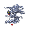
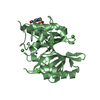
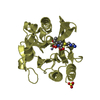
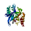
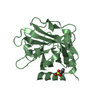
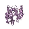
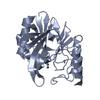
- PDB-1elt: STRUCTURE OF NATIVE PANCREATIC ELASTASE FROM NORTH ATLANTIC SALMO... -
+ Open data

- Basic information
| Entry | Database: PDB / ID: 1elt | ||||||
|---|---|---|---|---|---|---|---|
| Title | STRUCTURE OF NATIVE PANCREATIC ELASTASE FROM NORTH ATLANTIC SALMON AT 1.61 ANGSTROMS RESOLUTION | ||||||
 Components Components | ELASTASE | ||||||
 Keywords Keywords | SERINE PROTEINASE | ||||||
| Function / homology |  Function and homology information Function and homology informationpancreatic elastase / serine-type endopeptidase activity / proteolysis / : / metal ion binding Similarity search - Function | ||||||
| Biological species |  | ||||||
| Method |  X-RAY DIFFRACTION / Resolution: 1.61 Å X-RAY DIFFRACTION / Resolution: 1.61 Å | ||||||
 Authors Authors | Berglund, G.I. / Smalaas, A.O. | ||||||
 Citation Citation | Journal: Acta Crystallogr.,Sect.D / Year: 1995 Title: Structure of native pancreatic elastase from North Atlantic salmon at 1.61 A resolution. Authors: Berglund, G.I. / Willassen, N.P. / Hordvik, A. / Smalas, A.O. #1: Journal: To be PublishedTitle: Crystallization and Preliminary X-Ray Crystallographic Studies of Native Elastase from North Atlantic Salmon (Salmo Salar) Authors: Berglund, G.I. / Smalaas, A.O. / Hansen, L.Kr. / Willassen, N.P. #2: Journal: Biochemistry / Year: 1989Title: Human Leukocyte and Porcine Pancreatic Elastase: X-Ray Crystal Structures, Mechanism, Substrate Specificity, and Mechanism-Based Inhibitors Authors: Bode, W. / Meyer, E. / Powers, J.C. #3: Journal: Acta Crystallogr.,Sect.B / Year: 1988Title: Structure of Native Porcine Pancreatic Elastase at 1.65 Angstroms Resolution Authors: Meyer, E. / Cole, G. / Radhakrishnan, R. | ||||||
| History |
|
- Structure visualization
| Structure viewer | Molecule: MolmilJmol/JSmol |
|---|
- Downloads & links
-Download
| PDBx/mmCIF format | 1elt.cif.gz | 60.3 KB | Display | PDBx/mmCIF format |
|---|---|---|---|---|
| PDB format | pdb1elt.ent.gz | 43.4 KB | Display | PDB format |
| PDBx/mmJSON format | 1elt.json.gz | Tree view | PDBx/mmJSON format | |
| Others |  Other downloads Other downloads |
-Validation report
| Arichive directory | https://data.pdbj.org/pub/pdb/validation_reports/el/1eltftp://data.pdbj.org/pub/pdb/validation_reports/el/1elt | HTTPS FTP |
|---|
-Related structure data
| Similar structure data |
|---|


-Links
 PDBj
PDBj
- Assembly
| Deposited unit | 
| |||||||||
|---|---|---|---|---|---|---|---|---|---|---|
| 1 |
| |||||||||
| Unit cell |
| |||||||||
| Components on special symmetry positions |
|
-Components
| #1: Protein | Mass: 25033.883 Da / Num. of mol.: 1 / Source method: isolated from a natural source / Source: (natural) | ||
|---|---|---|---|
| #2: Chemical | ChemComp-CA /   Mass: 40.078 Da / Num. of mol.: 1 / Source method: obtained synthetically / Formula: Ca Mass: 40.078 Da / Num. of mol.: 1 / Source method: obtained synthetically / Formula: Ca | ||
| #3: Water | ChemComp-HOH /  Mass: 18.015 Da / Num. of mol.: 156 / Source method: isolated from a natural source / Formula: H2O Mass: 18.015 Da / Num. of mol.: 156 / Source method: isolated from a natural source / Formula: H2O | ||
| Has protein modification | Y | ||
| Nonpolymer details | CALCIUM IS STRUCTURAL| Sequence details | THE AMINO ACID NUMBERING SYSTEM IS THE ONE ADOPTED FROM CHYMOTRYPS | |
-Experimental details
-Experiment
| Experiment | Method: X-RAY DIFFRACTION |
|---|
- Sample preparation
| Crystal | Density Matthews: 1.94 Å3/Da / Density % sol: 36.54 % | ||||||||||||||||||||||||||||||||||||||||
|---|---|---|---|---|---|---|---|---|---|---|---|---|---|---|---|---|---|---|---|---|---|---|---|---|---|---|---|---|---|---|---|---|---|---|---|---|---|---|---|---|---|
| Crystal grow | *PLUS pH: 7.3 / Method: vapor diffusion, hanging drop | ||||||||||||||||||||||||||||||||||||||||
| Components of the solutions | *PLUS
|
-Data collection
| Diffraction source | Wavelength: 1.5418 |
|---|---|
| Detector | Type: ENRAF-NONIUS FAST / Detector: DIFFRACTOMETER / Date: Jul 8, 1993 |
| Radiation | Monochromatic (M) / Laue (L): M / Scattering type: x-ray |
| Radiation wavelength | Wavelength: 1.5418 Å / Relative weight: 1 |
| Reflection | Num. obs: 21574 / % possible obs: 83.6 % / Redundancy: 1.7 % / Rmerge(I) obs: 0.024 |
| Reflection | *PLUS Highest resolution: 1.61 Å / Lowest resolution: 4.82 Å / Num. measured all: 36833 / Rmerge(I) obs: 0.024 |
| Reflection shell | *PLUS Highest resolution: 1.61 Å / Lowest resolution: 1.71 Å / % possible obs: 46.1 % / Redundancy: 1.2 % / Num. unique obs: 1660 / Num. measured obs: 2002 / Rmerge(I) obs: 0.174 / Mean I/σ(I) obs: 60.8 |
- Processing
| Software |
| |||||||||||||||||||||||||||||||||||||||||||||||||||||||||||||||
|---|---|---|---|---|---|---|---|---|---|---|---|---|---|---|---|---|---|---|---|---|---|---|---|---|---|---|---|---|---|---|---|---|---|---|---|---|---|---|---|---|---|---|---|---|---|---|---|---|---|---|---|---|---|---|---|---|---|---|---|---|---|---|---|---|
| Refinement | Resolution: 1.61→8 Å / σ(F): 0
| |||||||||||||||||||||||||||||||||||||||||||||||||||||||||||||||
| Displacement parameters | Biso mean: 14.6 Å2 | |||||||||||||||||||||||||||||||||||||||||||||||||||||||||||||||
| Refine analyze | Luzzati coordinate error obs: 0.15 Å | |||||||||||||||||||||||||||||||||||||||||||||||||||||||||||||||
| Refinement step | Cycle: LAST / Resolution: 1.61→8 Å
| |||||||||||||||||||||||||||||||||||||||||||||||||||||||||||||||
| Refine LS restraints |
| |||||||||||||||||||||||||||||||||||||||||||||||||||||||||||||||
| Refine LS restraints | *PLUS
|
