 Movie
Movie Controller

[English] 日本語
 Yorodumi
- PDB-1eis: UDA UNCOMPLEXED FORM. CRYSTAL STRUCTURE OF URTICA DIOICA AGGLUTIN... -
+ Open data

- Basic information
| Entry | Database: PDB / ID: 1eis | |||||||||
|---|---|---|---|---|---|---|---|---|---|---|
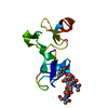
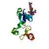
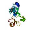
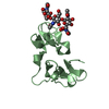
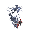
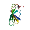
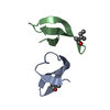
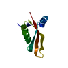
| Title | UDA UNCOMPLEXED FORM. CRYSTAL STRUCTURE OF URTICA DIOICA AGGLUTININ, A SUPERANTIGEN PRESENTED BY MHC MOLECULES OF CLASS I AND CLASS II | |||||||||
 Components Components | PROTEIN (AGGLUTININ ISOLECTIN VI/AGGLUTININ ISOLECTIN V) | |||||||||
 Keywords Keywords | SUGAR BINDING PROTEIN / LECTIN / HEVEIN DOMAIN / UDA / SUPERANTIGEN | |||||||||
| Function / homology | Chitin-binding, type 1, conserved site / Chitin recognition protein / Chitin recognition or binding domain signature. / Chitin-binding type-1 domain profile. / Chitin binding domain / Chitin-binding, type 1 / Endochitinase-like superfamily / chitin binding / Agglutinin isolectin VI Function and homology information Function and homology information | |||||||||
| Biological species |  Urtica dioica (great nettle) Urtica dioica (great nettle) | |||||||||
| Method |  X-RAY DIFFRACTION / SYNCHROTRON / MIR / Resolution: 1.66 Å X-RAY DIFFRACTION / SYNCHROTRON / MIR / Resolution: 1.66 Å | |||||||||
 Authors Authors | Saul, F.A. / Rovira, P. / Boulot, G. / Van Damme, E.J.M. / Peumans, W.J. / Truffa-Bachi, P. / Bentley, G.A. | |||||||||
 Citation Citation | Journal: Structure Fold.Des. / Year: 2000 Title: Crystal structure of Urtica dioica agglutinin, a superantigen presented by MHC molecules of class I and class II. Authors: Saul, F.A. / Rovira, P. / Boulot, G. / Damme, E.J. / Peumans, W.J. / Truffa-Bachi, P. / Bentley, G.A. #1: Journal: Plant Mol.Biol. / Year: 1999Title: Characterisation of Urtica dioica Agglutinin Isolectins and the Encoding Gene Family Authors: Does, M.P. / Ng, D.K. / Dekker, H.L. / Peumans, W.J. / Houterman, P.M. / Van Damme, E.J. / Cornelissen, B.J.C. | |||||||||
| History |
|
- Structure visualization
| Structure viewer | Molecule: MolmilJmol/JSmol |
|---|
- Downloads & links
-Download
| PDBx/mmCIF format | 1eis.cif.gz | 30.1 KB | Display | PDBx/mmCIF format |
|---|---|---|---|---|
| PDB format | pdb1eis.ent.gz | 18.9 KB | Display | PDB format |
| PDBx/mmJSON format | 1eis.json.gz | Tree view | PDBx/mmJSON format | |
| Others |  Other downloads Other downloads |
-Validation report
| Arichive directory | https://data.pdbj.org/pub/pdb/validation_reports/ei/1eisftp://data.pdbj.org/pub/pdb/validation_reports/ei/1eis | HTTPS FTP |
|---|
-Related structure data

-Links
 PDBj
PDBj- Assembly
| Deposited unit | 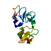
| ||||||||
|---|---|---|---|---|---|---|---|---|---|
| 1 |
| ||||||||
| Unit cell |
|
-Components
| #1: Protein | Mass: 9350.269 Da / Num. of mol.: 1 / Source method: isolated from a natural source / Details: PURIFIED FROM THE RHIZOMES / Source: (natural) Urtica dioica (great nettle) / References: UniProt: Q9SYR5 |
|---|---|
| #2: Water | ChemComp-HOH /  Mass: 18.015 Da / Num. of mol.: 49 / Source method: isolated from a natural source / Formula: H2O Mass: 18.015 Da / Num. of mol.: 49 / Source method: isolated from a natural source / Formula: H2O |
| Compound details | The structure comprises two hevein-like domains, each homologous to those of Wheat Germ Agglutinin ...The structure comprises two hevein-like domains, each homologous to those of Wheat Germ Agglutinin (PDB code: 9wga). Each domain contains a saccharide-binding site. The N-terminal residue is pyrrolidone carboxylic acid (PCA). A dual conformation is seen at Gly79, Gly80. No electron density is seen for the side chain of Arg33 beyond the CB atom. No interpretable density is seen for C-terminal residues Ser87, Ser88, and Ser89. |
| Has protein modification | Y |
-Experimental details
-Experiment
| Experiment | Method: X-RAY DIFFRACTION / Number of used crystals: 3 |
|---|
- Sample preparation
| Crystal | Density Matthews: 2.68 Å3/Da / Density % sol: 54.16 % | |||||||||||||||||||||||||
|---|---|---|---|---|---|---|---|---|---|---|---|---|---|---|---|---|---|---|---|---|---|---|---|---|---|---|
| Crystal grow | Temperature: 290 K / Method: vapor diffusion, sitting drop / pH: 6 Details: PEG 6000, sodium acetate, sodium chloride , pH 6.0, VAPOR DIFFUSION, SITTING DROP, temperature 290.0K | |||||||||||||||||||||||||
| Crystal grow | *PLUS PH range low: 6.5 / PH range high: 5.5 | |||||||||||||||||||||||||
| Components of the solutions | *PLUS
|
-Data collection
| Diffraction | Mean temperature: 298 K |
|---|---|
| Diffraction source | Source: SYNCHROTRON / Site: LURE  / Beamline: D41A / Wavelength: 1.375 / Beamline: D41A / Wavelength: 1.375 |
| Detector | Type: MARRESEARCH / Detector: IMAGE PLATE / Date: Sep 21, 1998 |
| Radiation | Protocol: SINGLE WAVELENGTH / Monochromatic (M) / Laue (L): M / Scattering type: x-ray |
| Radiation wavelength | Wavelength: 1.375 Å / Relative weight: 1 |
| Reflection | Resolution: 1.66→18 Å / Num. all: 11689 / Num. obs: 11689 / % possible obs: 95.4 % / Observed criterion σ(F): 0 / Observed criterion σ(I): 0 / Redundancy: 12.5 % / Biso Wilson estimate: 30.8 Å2 / Rmerge(I) obs: 0.056 / Net I/σ(I): 24.7 |
| Reflection shell | Resolution: 1.66→1.72 Å / Redundancy: 8.9 % / Rmerge(I) obs: 0.352 / Num. unique all: 1015 / % possible all: 84.7 |
| Reflection shell | *PLUS % possible obs: 84.6 % |
- Processing
| Software |
| ||||||||||||||||||||||||||||||||||||||||||||||||||||||||||||||||
|---|---|---|---|---|---|---|---|---|---|---|---|---|---|---|---|---|---|---|---|---|---|---|---|---|---|---|---|---|---|---|---|---|---|---|---|---|---|---|---|---|---|---|---|---|---|---|---|---|---|---|---|---|---|---|---|---|---|---|---|---|---|---|---|---|---|
| Refinement | Method to determine structure: MIR / Resolution: 1.66→18 Å / σ(F): 0 / σ(I): 0 / Stereochemistry target values: Engh & Huber Details: The structure was determined to 2.45A resolution by MIR methods based on 4 Hg derivatives (rotating-anode X-ray source) with solvent flattening, and refined to 1.66A resolution with ...Details: The structure was determined to 2.45A resolution by MIR methods based on 4 Hg derivatives (rotating-anode X-ray source) with solvent flattening, and refined to 1.66A resolution with synchrotron data using a bulk solvent correction and anisotropic scale factor.
| ||||||||||||||||||||||||||||||||||||||||||||||||||||||||||||||||
| Refinement step | Cycle: LAST / Resolution: 1.66→18 Å
| ||||||||||||||||||||||||||||||||||||||||||||||||||||||||||||||||
| Refine LS restraints |
|
