[English] 日本語
 Yorodumi
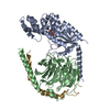
- PDB-1ei5: CRYSTAL STRUCTURE OF A D-AMINOPEPTIDASE FROM OCHROBACTRUM ANTHROPI -
+ Open data
- Basic information
| Entry | Database: PDB / ID: 1ei5 | ||||||
|---|---|---|---|---|---|---|---|
| Title | CRYSTAL STRUCTURE OF A D-AMINOPEPTIDASE FROM OCHROBACTRUM ANTHROPI | ||||||
 Components Components | D-AMINOPEPTIDASE | ||||||
 Keywords Keywords | HYDROLASE / D-AMINOPEPTIDASE / PENICILLIN BINDING PROTEIN / ALPHA/BETA DOMAIN / BETA BARREL DOMAIN | ||||||
| Function / homology |  Function and homology information Function and homology information | ||||||
| Biological species |  Ochrobactrum anthropi (bacteria) Ochrobactrum anthropi (bacteria) | ||||||
| Method |  X-RAY DIFFRACTION / X-RAY DIFFRACTION /  SYNCHROTRON / Resolution: 1.9 Å SYNCHROTRON / Resolution: 1.9 Å | ||||||
 Authors Authors | Bompard-Gilles, C. / Remaut, H. / Villeret, V. / Prange, T. / Fanuel, L. / Joris, J. / Frere, J.-M. / Van Beeumen, J. | ||||||
 Citation Citation |  Journal: Structure Fold.Des. / Year: 2000 Journal: Structure Fold.Des. / Year: 2000Title: Crystal structure of a D-aminopeptidase from Ochrobactrum anthropi, a new member of the 'penicillin-recognizing enzyme' family. Authors: Bompard-Gilles, C. / Remaut, H. / Villeret, V. / Prange, T. / Fanuel, L. / Delmarcelle, M. / Joris, B. / Frere, J. / Van Beeumen, J. | ||||||
| History |
|
- Structure visualization
| Structure viewer | Molecule:  Molmil Molmil Jmol/JSmol Jmol/JSmol |
|---|
- Downloads & links
- Download
| PDBx/mmCIF format |  1ei5.cif.gz 1ei5.cif.gz | 118.2 KB | Display |  PDBx/mmCIF format PDBx/mmCIF format |
|---|---|---|---|---|
| PDB format |  pdb1ei5.ent.gz pdb1ei5.ent.gz | 90 KB | Display |  PDB format PDB format |
| PDBx/mmJSON format |  1ei5.json.gz 1ei5.json.gz | Tree view |  PDBx/mmJSON format PDBx/mmJSON format | |
| Others |  Other downloads Other downloads |
-Validation report
| Arichive directory |  https://data.pdbj.org/pub/pdb/validation_reports/ei/1ei5 https://data.pdbj.org/pub/pdb/validation_reports/ei/1ei5 ftp://data.pdbj.org/pub/pdb/validation_reports/ei/1ei5 ftp://data.pdbj.org/pub/pdb/validation_reports/ei/1ei5 | HTTPS FTP |
|---|
-Related structure data
| Similar structure data |
|---|
- Links
Links
- Assembly
| Deposited unit | 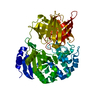
| ||||||||
|---|---|---|---|---|---|---|---|---|---|
| 1 | 
| ||||||||
| Unit cell |
| ||||||||
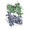
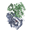
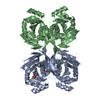
| Details | The biological assembly is a dimer constructed from chain A a symmetry partner generated by the two-fold. |
- Components
| #1: Protein | Mass: 57458.754 Da / Num. of mol.: 1 Source method: isolated from a genetically manipulated source Source: (gene. exp.)  Ochrobactrum anthropi (bacteria) / Strain: SCRC C1-38 / Plasmid: PUC18 / Production host: Ochrobactrum anthropi (bacteria) / Strain: SCRC C1-38 / Plasmid: PUC18 / Production host:  References: UniProt: Q9ZBA9, D-stereospecific aminopeptidase |
|---|---|
| #2: Water | ChemComp-HOH / |
-Experimental details
-Experiment
| Experiment | Method:  X-RAY DIFFRACTION / Number of used crystals: 1 X-RAY DIFFRACTION / Number of used crystals: 1 |
|---|
- Sample preparation
| Crystal | Density Matthews: 3.06 Å3/Da / Density % sol: 59.75 % | ||||||||||||||||||||||||||||||||||||||||
|---|---|---|---|---|---|---|---|---|---|---|---|---|---|---|---|---|---|---|---|---|---|---|---|---|---|---|---|---|---|---|---|---|---|---|---|---|---|---|---|---|---|
| Crystal grow | Temperature: 294 K / Method: vapor diffusion, hanging drop / pH: 5 Details: PEG 400, sodium acetate, pH 5, VAPOR DIFFUSION, HANGING DROP, temperature 294K | ||||||||||||||||||||||||||||||||||||||||
| Crystal grow | *PLUS Temperature: 21 ℃ / pH: 8 Details: drop consists of equal amounts of protein and reservoir solutions | ||||||||||||||||||||||||||||||||||||||||
| Components of the solutions | *PLUS
|
-Data collection
| Diffraction | Mean temperature: 277 K |
|---|---|
| Diffraction source | Source:  SYNCHROTRON / Site: LURE SYNCHROTRON / Site: LURE  / Beamline: DW32 / Wavelength: 0.97 / Beamline: DW32 / Wavelength: 0.97 |
| Detector | Type: MARRESEARCH / Detector: IMAGE PLATE / Date: Oct 4, 1998 |
| Radiation | Protocol: SINGLE WAVELENGTH / Monochromatic (M) / Laue (L): M / Scattering type: x-ray |
| Radiation wavelength | Wavelength: 0.97 Å / Relative weight: 1 |
| Reflection | Resolution: 1.82→15 Å / Num. all: 62212 / Num. obs: 62212 / % possible obs: 96.5 % / Observed criterion σ(F): 0 / Observed criterion σ(I): 0 / Redundancy: 12.99 % / Biso Wilson estimate: 17.93 Å2 / Rmerge(I) obs: 0.092 / Net I/σ(I): 14.6 |
| Reflection shell | Resolution: 1.82→1.89 Å / Redundancy: 4.76 % / Rmerge(I) obs: 0.346 / Num. unique all: 5161 / % possible all: 81 |
| Reflection | *PLUS % possible obs: 93.2 % / Redundancy: 7.9 % / Rmerge(I) obs: 0.052 |
| Reflection shell | *PLUS % possible obs: 81 % / Mean I/σ(I) obs: 3.5 |
- Processing
| Software |
| ||||||||||||||||||||
|---|---|---|---|---|---|---|---|---|---|---|---|---|---|---|---|---|---|---|---|---|---|
| Refinement | Resolution: 1.9→12.5 Å / σ(F): 0 / σ(I): 0 / Stereochemistry target values: Engh & Huber
| ||||||||||||||||||||
| Refinement step | Cycle: LAST / Resolution: 1.9→12.5 Å
| ||||||||||||||||||||
| Refine LS restraints |
| ||||||||||||||||||||
| Software | *PLUS Name: REFMAC / Classification: refinement | ||||||||||||||||||||
| Refinement | *PLUS Num. reflection all: 54802 | ||||||||||||||||||||
| Solvent computation | *PLUS | ||||||||||||||||||||
| Displacement parameters | *PLUS | ||||||||||||||||||||
| Refine LS restraints | *PLUS Type: p_angle_d / Dev ideal: 0.031 |
 Movie
Movie Controller



 PDBj
PDBj

