 ムービー
ムービー コントローラー


+ データを開く

- 基本情報
基本情報
| 登録情報 | データベース: PDB / ID: 1eg4 | ||||||
|---|---|---|---|---|---|---|---|
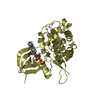
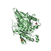
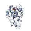
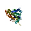
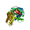
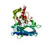
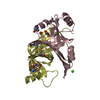
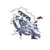
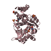
| タイトル | STRUCTURE OF A DYSTROPHIN WW DOMAIN FRAGMENT IN COMPLEX WITH A BETA-DYSTROGLYCAN PEPTIDE | ||||||
 要素 要素 |
| ||||||
 キーワード キーワード | STRUCTURAL PROTEIN / EF-hand like domain / WW domain / Polyproline type II (PPII) helix | ||||||
| 機能・相同性 |  機能・相同性情報 機能・相同性情報Defective POMT2 causes MDDGA2, MDDGB2 and MDDGC2 / Defective POMT1 causes MDDGA1, MDDGB1 and MDDGC1 / muscle attachment / dystroglycan complex / nerve maturation / Defective POMGNT1 causes MDDGA3, MDDGB3 and MDDGC3 / retrograde trans-synaptic signaling by trans-synaptic protein complex / regulation of muscle system process / O-linked glycosylation / regulation of cellular response to growth factor stimulus ...Defective POMT2 causes MDDGA2, MDDGB2 and MDDGC2 / Defective POMT1 causes MDDGA1, MDDGB1 and MDDGC1 / muscle attachment / dystroglycan complex / nerve maturation / Defective POMGNT1 causes MDDGA3, MDDGB3 and MDDGC3 / retrograde trans-synaptic signaling by trans-synaptic protein complex / regulation of muscle system process / O-linked glycosylation / regulation of cellular response to growth factor stimulus / syntrophin complex / contractile ring / cardiac muscle cell action potential / calcium-dependent cell-matrix adhesion / morphogenesis of an epithelial sheet / regulation of skeletal muscle contraction / microtubule anchoring / synaptic signaling / dystrophin-associated glycoprotein complex / laminin-1 binding / response to denervation involved in regulation of muscle adaptation / cell-substrate junction / peptide biosynthetic process / motile cilium assembly / photoreceptor ribbon synapse / nerve development / dystroglycan binding / basement membrane organization / regulation of skeletal muscle contraction by regulation of release of sequestered calcium ion / positive regulation of myelination / cellular response to cholesterol / vinculin binding / EGR2 and SOX10-mediated initiation of Schwann cell myelination / regulation of sodium ion transmembrane transport / branching involved in salivary gland morphogenesis / Formation of the dystrophin-glycoprotein complex (DGC) / skeletal muscle tissue regeneration / myelination in peripheral nervous system / costamere / commissural neuron axon guidance / muscle cell development / regulation of calcium ion transmembrane transport / neuron projection terminus / node of Ranvier / Striated Muscle Contraction / filopodium membrane / angiogenesis involved in wound healing / axon regeneration / structural constituent of muscle / positive regulation of cell-matrix adhesion / muscle organ development / muscle cell cellular homeostasis / regulation of synapse organization / myosin binding / response to muscle activity / epithelial tube branching involved in lung morphogenesis / maintenance of blood-brain barrier / nitric-oxide synthase binding / regulation of neurotransmitter receptor localization to postsynaptic specialization membrane / alpha-actinin binding / membrane protein ectodomain proteolysis / positive regulation of oligodendrocyte differentiation / basement membrane / Non-integrin membrane-ECM interactions / plasma membrane raft / ECM proteoglycans / neuron development / postsynaptic cytosol / regulation of cardiac muscle contraction by regulation of the release of sequestered calcium ion / heart morphogenesis / skeletal muscle tissue development / negative regulation of MAPK cascade / cardiac muscle contraction / laminin binding / regulation of release of sequestered calcium ion into cytosol by sarcoplasmic reticulum / response to muscle stretch / extracellular matrix organization / positive regulation of neuron differentiation / negative regulation of phosphatidylinositol 3-kinase/protein kinase B signal transduction / tubulin binding / SH2 domain binding / nuclear periphery / regulation of heart rate / axon guidance / negative regulation of cell migration / morphogenesis of an epithelium / adherens junction / filopodium / cellular response to mechanical stimulus / response to peptide hormone / positive regulation of neuron projection development / sarcolemma / regulation of synaptic plasticity / GABA-ergic synapse / structural constituent of cytoskeleton / Golgi lumen / Regulation of expression of SLITs and ROBOs / Z disc / intracellular protein localization / protein transport 類似検索 - 分子機能 | ||||||
| 生物種 |  Homo sapiens (ヒト) Homo sapiens (ヒト) | ||||||
| 手法 |  X線回折 / シンクロトロン / 解像度: 2 Å X線回折 / シンクロトロン / 解像度: 2 Å | ||||||
 データ登録者 データ登録者 | Huang, X. / Poy, F. / Zhang, R. / Joachimiak, A. / Sudol, M. / Eck, M.J. | ||||||
 引用 引用 | ジャーナル: Nat.Struct.Biol. / 年: 2000 タイトル: Structure of a WW domain containing fragment of dystrophin in complex with beta-dystroglycan. 著者: Huang, X. / Poy, F. / Zhang, R. / Joachimiak, A. / Sudol, M. / Eck, M.J. | ||||||
| 履歴 |
|
- 構造の表示
構造の表示
| 構造ビューア | 分子: MolmilJmol/JSmol |
|---|
- ダウンロードとリンク
-ダウンロード
| PDBx/mmCIF形式 | 1eg4.cif.gz | 73.3 KB | 表示 | PDBx/mmCIF形式 |
|---|---|---|---|---|
| PDB形式 | pdb1eg4.ent.gz | 53.8 KB | 表示 | PDB形式 |
| PDBx/mmJSON形式 | 1eg4.json.gz | ツリー表示 | PDBx/mmJSON形式 | |
| その他 |  その他のダウンロード その他のダウンロード |
-検証レポート
| 文書・要旨 | 1eg4_validation.pdf.gz | 372.4 KB | 表示 | wwPDB検証レポート |
|---|---|---|---|---|
| 文書・詳細版 | 1eg4_full_validation.pdf.gz | 381.8 KB | 表示 | |
| XML形式データ | 1eg4_validation.xml.gz | 8.2 KB | 表示 | |
| CIF形式データ | 1eg4_validation.cif.gz | 13.3 KB | 表示 | |
| アーカイブディレクトリ | https://data.pdbj.org/pub/pdb/validation_reports/eg/1eg4ftp://data.pdbj.org/pub/pdb/validation_reports/eg/1eg4 | HTTPS FTP |
-関連構造データ


-リンク
 PDBj
PDBj


- 集合体
集合体
| 登録構造単位 | 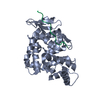
| ||||||||
|---|---|---|---|---|---|---|---|---|---|
| 1 |
| ||||||||
| 単位格子 |
|
-要素
| #1: タンパク質・ペプチド | 分子量: 1746.036 Da / 分子数: 1 / 由来タイプ: 合成 詳細: This peptide was chemically synthesized and occurs naturally in Homo Sapiens (Human) 参照: UniProt: Q14118 |
|---|---|
| #2: タンパク質 | 分子量: 29845.232 Da / 分子数: 1 / Fragment: WW DOMAIN / 由来タイプ: 組換発現 / 由来: (組換発現) Homo sapiens (ヒト) / プラスミド: PGEX-2TK / 発現宿主:  |
| #3: 水 | ChemComp-HOH /  分子量: 18.015 Da / 分子数: 283 / 由来タイプ: 天然 / 式: H2O 分子量: 18.015 Da / 分子数: 283 / 由来タイプ: 天然 / 式: H2O |
-実験情報
-実験
| 実験 | 手法: X線回折 / 使用した結晶の数: 2 |
|---|
- 試料調製
試料調製
| 結晶 | マシュー密度: 2.16 Å3/Da / 溶媒含有率: 43.17 % | ||||||||||||||||||||||||||||||||||||||||||||||||||||||
|---|---|---|---|---|---|---|---|---|---|---|---|---|---|---|---|---|---|---|---|---|---|---|---|---|---|---|---|---|---|---|---|---|---|---|---|---|---|---|---|---|---|---|---|---|---|---|---|---|---|---|---|---|---|---|---|
| 結晶化 | 温度: 298 K / 手法: 蒸気拡散法, ハンギングドロップ法 / pH: 7 詳細: ammonium sulfate, glycerol, DTT, pH 7.0, VAPOR DIFFUSION, HANGING DROP, temperature 298K | ||||||||||||||||||||||||||||||||||||||||||||||||||||||
| 結晶化 | *PLUS 温度: 22 ℃ / pH: 6.5 | ||||||||||||||||||||||||||||||||||||||||||||||||||||||
| 溶液の組成 | *PLUS
|
-データ収集
| 回折 | 平均測定温度: 213 K |
|---|---|
| 放射光源 | 由来: シンクロトロン / サイト: APS  / ビームライン: 19-ID / 波長: 1.07 / ビームライン: 19-ID / 波長: 1.07 |
| 検出器 | タイプ: APS-1 / 検出器: CCD / 日付: 1999年4月12日 |
| 放射 | プロトコル: SINGLE WAVELENGTH / 単色(M)・ラウエ(L): M / 散乱光タイプ: x-ray |
| 放射波長 | 波長: 1.07 Å / 相対比: 1 |
| 反射 | 解像度: 1.9→25 Å / Num. all: 120237 / Num. obs: 21895 / % possible obs: 98.3 % / Observed criterion σ(I): -3 / 冗長度: 5.5 % / Biso Wilson estimate: 24 Å2 / Rmerge(I) obs: 0.052 / Net I/σ(I): 12.6 |
| 反射 シェル | 解像度: 1.9→1.97 Å / 冗長度: 5 % / Rmerge(I) obs: 0.295 / Num. unique all: 2146 / % possible all: 95.3 |
| 反射 | *PLUS Num. measured all: 120237 |
- 解析
解析
| ソフトウェア |
| |||||||||||||||||||||||||
|---|---|---|---|---|---|---|---|---|---|---|---|---|---|---|---|---|---|---|---|---|---|---|---|---|---|---|
| 精密化 | 解像度: 2→20 Å / σ(F): 2 / 立体化学のターゲット値: Engh & Huber
| |||||||||||||||||||||||||
| 精密化ステップ | サイクル: LAST / 解像度: 2→20 Å
| |||||||||||||||||||||||||
| 拘束条件 |
| |||||||||||||||||||||||||
| ソフトウェア | *PLUS 名称: X-PLOR / バージョン: 3.1 / 分類: refinement | |||||||||||||||||||||||||
| 精密化 | *PLUS 最高解像度: 2 Å / 最低解像度: 20 Å / σ(F): 2 / Rfactor obs: 0.197 | |||||||||||||||||||||||||
| 溶媒の処理 | *PLUS | |||||||||||||||||||||||||
| 原子変位パラメータ | *PLUS |
