 Movie
Movie Controller

[English] 日本語
 Yorodumi
- PDB-1eg4: STRUCTURE OF A DYSTROPHIN WW DOMAIN FRAGMENT IN COMPLEX WITH A BE... -
+ Open data

- Basic information
| Entry | Database: PDB / ID: 1eg4 | ||||||
|---|---|---|---|---|---|---|---|
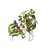
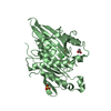
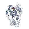
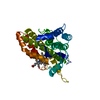
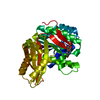
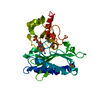
| Title | STRUCTURE OF A DYSTROPHIN WW DOMAIN FRAGMENT IN COMPLEX WITH A BETA-DYSTROGLYCAN PEPTIDE | ||||||
 Components Components |
| ||||||
 Keywords Keywords | STRUCTURAL PROTEIN / EF-hand like domain / WW domain / Polyproline type II (PPII) helix | ||||||
| Function / homology |  Function and homology information Function and homology informationDefective POMT2 causes MDDGA2, MDDGB2 and MDDGC2 / Defective POMT1 causes MDDGA1, MDDGB1 and MDDGC1 / DAG1 core M3 glycosylations / dystroglycan complex / nerve maturation / Defective POMGNT1 causes MDDGA3, MDDGB3 and MDDGC3 / DAG1 core M2 glycosylations / DAG1 core M1 glycosylations / muscle attachment / retrograde trans-synaptic signaling by trans-synaptic protein complex ...Defective POMT2 causes MDDGA2, MDDGB2 and MDDGC2 / Defective POMT1 causes MDDGA1, MDDGB1 and MDDGC1 / DAG1 core M3 glycosylations / dystroglycan complex / nerve maturation / Defective POMGNT1 causes MDDGA3, MDDGB3 and MDDGC3 / DAG1 core M2 glycosylations / DAG1 core M1 glycosylations / muscle attachment / retrograde trans-synaptic signaling by trans-synaptic protein complex / regulation of muscle system process / regulation of cellular response to growth factor stimulus / syntrophin complex / Matriglycan biosynthesis on DAG1 / morphogenesis of an epithelial sheet / contractile ring / calcium-dependent cell-matrix adhesion / cardiac muscle cell action potential / microtubule anchoring / synaptic signaling / dystrophin-associated glycoprotein complex / laminin-1 binding / response to denervation involved in regulation of muscle adaptation / cell-substrate junction / branching involved in salivary gland morphogenesis / basement membrane organization / motile cilium assembly / peptide biosynthetic process / photoreceptor ribbon synapse / nerve development / dystroglycan binding / regulation of skeletal muscle contraction by regulation of release of sequestered calcium ion / positive regulation of myelination / vinculin binding / myelination in peripheral nervous system / cellular response to cholesterol / camera-type eye development / EGR2 and SOX10-mediated initiation of Schwann cell myelination / skeletal muscle tissue regeneration / regulation of sodium ion transmembrane transport / costamere / Formation of the dystrophin-glycoprotein complex (DGC) / muscle cell development / commissural neuron axon guidance / angiogenesis involved in wound healing / regulation of calcium ion transmembrane transport / Striated Muscle Contraction / node of Ranvier / muscle cell cellular homeostasis / filopodium membrane / axon regeneration / inhibitory synapse assembly / muscle organ development / response to muscle activity / positive regulation of cell-matrix adhesion / structural constituent of muscle / positive regulation of oligodendrocyte differentiation / maintenance of blood-brain barrier / myosin binding / epithelial tube branching involved in lung morphogenesis / regulation of synapse organization / laminin receptor activity / neuron projection terminus / regulation of neurotransmitter receptor localization to postsynaptic specialization membrane / membrane protein ectodomain proteolysis / nitric-oxide synthase binding / basement membrane / regulation of skeletal muscle contraction / skeletal muscle tissue development / alpha-actinin binding / Non-integrin membrane-ECM interactions / heart morphogenesis / neuron development / plasma membrane raft / ECM proteoglycans / postsynaptic cytosol / response to muscle stretch / positive regulation of Rac protein signal transduction / cardiac muscle contraction / negative regulation of MAPK cascade / extracellular matrix organization / regulation of release of sequestered calcium ion into cytosol by sarcoplasmic reticulum / regulation of cardiac muscle contraction by regulation of the release of sequestered calcium ion / positive regulation of neuron differentiation / laminin binding / regulation of heart rate / nuclear periphery / axon guidance / SH2 domain binding / negative regulation of cell migration / morphogenesis of an epithelium / negative regulation of phosphatidylinositol 3-kinase/protein kinase B signal transduction / filopodium / adherens junction / positive regulation of neuron projection development / cellular response to mechanical stimulus / tubulin binding / regulation of synaptic plasticity / sarcolemma / Golgi lumen Similarity search - Function | ||||||
| Biological species |  Homo sapiens (human) Homo sapiens (human) | ||||||
| Method |  X-RAY DIFFRACTION / SYNCHROTRON / Resolution: 2 Å X-RAY DIFFRACTION / SYNCHROTRON / Resolution: 2 Å | ||||||
 Authors Authors | Huang, X. / Poy, F. / Zhang, R. / Joachimiak, A. / Sudol, M. / Eck, M.J. | ||||||
 Citation Citation | Journal: Nat.Struct.Biol. / Year: 2000 Title: Structure of a WW domain containing fragment of dystrophin in complex with beta-dystroglycan. Authors: Huang, X. / Poy, F. / Zhang, R. / Joachimiak, A. / Sudol, M. / Eck, M.J. | ||||||
| History |
|
- Structure visualization
| Structure viewer | Molecule: MolmilJmol/JSmol |
|---|
- Downloads & links
-Download
| PDBx/mmCIF format | 1eg4.cif.gz | 73.3 KB | Display | PDBx/mmCIF format |
|---|---|---|---|---|
| PDB format | pdb1eg4.ent.gz | 53.8 KB | Display | PDB format |
| PDBx/mmJSON format | 1eg4.json.gz | Tree view | PDBx/mmJSON format | |
| Others |  Other downloads Other downloads |
-Validation report
| Arichive directory | https://data.pdbj.org/pub/pdb/validation_reports/eg/1eg4ftp://data.pdbj.org/pub/pdb/validation_reports/eg/1eg4 | HTTPS FTP |
|---|
-Related structure data


-Links
 PDBj
PDBj


- Assembly
| Deposited unit | 
| ||||||||
|---|---|---|---|---|---|---|---|---|---|
| 1 |
| ||||||||
| Unit cell |
|
-Components
| #1: Protein/peptide | Mass: 1746.036 Da / Num. of mol.: 1 / Source method: obtained synthetically Details: This peptide was chemically synthesized and occurs naturally in Homo Sapiens (Human) References: UniProt: Q14118 |
|---|---|
| #2: Protein | Mass: 29845.232 Da / Num. of mol.: 1 / Fragment: WW DOMAIN Source method: isolated from a genetically manipulated source Source: (gene. exp.) Homo sapiens (human) / Plasmid: PGEX-2TK / Production host:  |
| #3: Water | ChemComp-HOH /  Mass: 18.015 Da / Num. of mol.: 283 / Source method: isolated from a natural source / Formula: H2O Mass: 18.015 Da / Num. of mol.: 283 / Source method: isolated from a natural source / Formula: H2O |
-Experimental details
-Experiment
| Experiment | Method: X-RAY DIFFRACTION / Number of used crystals: 2 |
|---|
- Sample preparation
| Crystal | Density Matthews: 2.16 Å3/Da / Density % sol: 43.17 % | ||||||||||||||||||||||||||||||||||||||||||||||||||||||
|---|---|---|---|---|---|---|---|---|---|---|---|---|---|---|---|---|---|---|---|---|---|---|---|---|---|---|---|---|---|---|---|---|---|---|---|---|---|---|---|---|---|---|---|---|---|---|---|---|---|---|---|---|---|---|---|
| Crystal grow | Temperature: 298 K / Method: vapor diffusion, hanging drop / pH: 7 Details: ammonium sulfate, glycerol, DTT, pH 7.0, VAPOR DIFFUSION, HANGING DROP, temperature 298K | ||||||||||||||||||||||||||||||||||||||||||||||||||||||
| Crystal grow | *PLUS Temperature: 22 ℃ / pH: 6.5 | ||||||||||||||||||||||||||||||||||||||||||||||||||||||
| Components of the solutions | *PLUS
|
-Data collection
| Diffraction | Mean temperature: 213 K |
|---|---|
| Diffraction source | Source: SYNCHROTRON / Site: APS  / Beamline: 19-ID / Wavelength: 1.07 / Beamline: 19-ID / Wavelength: 1.07 |
| Detector | Type: APS-1 / Detector: CCD / Date: Apr 12, 1999 |
| Radiation | Protocol: SINGLE WAVELENGTH / Monochromatic (M) / Laue (L): M / Scattering type: x-ray |
| Radiation wavelength | Wavelength: 1.07 Å / Relative weight: 1 |
| Reflection | Resolution: 1.9→25 Å / Num. all: 120237 / Num. obs: 21895 / % possible obs: 98.3 % / Observed criterion σ(I): -3 / Redundancy: 5.5 % / Biso Wilson estimate: 24 Å2 / Rmerge(I) obs: 0.052 / Net I/σ(I): 12.6 |
| Reflection shell | Resolution: 1.9→1.97 Å / Redundancy: 5 % / Rmerge(I) obs: 0.295 / Num. unique all: 2146 / % possible all: 95.3 |
| Reflection | *PLUS Num. measured all: 120237 |
- Processing
| Software |
| |||||||||||||||||||||||||
|---|---|---|---|---|---|---|---|---|---|---|---|---|---|---|---|---|---|---|---|---|---|---|---|---|---|---|
| Refinement | Resolution: 2→20 Å / σ(F): 2 / Stereochemistry target values: Engh & Huber
| |||||||||||||||||||||||||
| Refinement step | Cycle: LAST / Resolution: 2→20 Å
| |||||||||||||||||||||||||
| Refine LS restraints |
| |||||||||||||||||||||||||
| Software | *PLUS Name: X-PLOR / Version: 3.1 / Classification: refinement | |||||||||||||||||||||||||
| Refinement | *PLUS Highest resolution: 2 Å / Lowest resolution: 20 Å / σ(F): 2 / Rfactor obs: 0.197 | |||||||||||||||||||||||||
| Solvent computation | *PLUS | |||||||||||||||||||||||||
| Displacement parameters | *PLUS |
