 Movie
Movie Controller

[English] 日本語
 Yorodumi
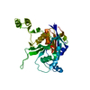
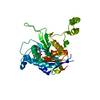
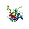
- PDB-1eg2: CRYSTAL STRUCTURE OF RHODOBACTER SPHEROIDES (N6 ADENOSINE) METHYL... -
+ Open data

- Basic information
| Entry | Database: PDB / ID: 1eg2 | ||||||
|---|---|---|---|---|---|---|---|
| Title | CRYSTAL STRUCTURE OF RHODOBACTER SPHEROIDES (N6 ADENOSINE) METHYLTRANSFERASE (M.RSRI) | ||||||
 Components Components | MODIFICATION METHYLASE RSRI | ||||||
 Keywords Keywords | TRANSFERASE / Rossmann Fold / exocyclic amino DNA methyltransferase RsrI / DNA binding / DNA modification / DNA methylation / Structural Genomics / PSI / Protein Structure Initiative / Midwest Center for Structural Genomics / MCSG | ||||||
| Function / homology |  Function and homology information Function and homology informationN-methyltransferase activity / site-specific DNA-methyltransferase (adenine-specific) / site-specific DNA-methyltransferase (adenine-specific) activity / DNA restriction-modification system / methylation / DNA binding Similarity search - Function | ||||||
| Biological species |  Rhodobacter sphaeroides (bacteria) Rhodobacter sphaeroides (bacteria) | ||||||
| Method |  X-RAY DIFFRACTION / SYNCHROTRON / Resolution: 1.75 Å X-RAY DIFFRACTION / SYNCHROTRON / Resolution: 1.75 Å | ||||||
 Authors Authors | Scavetta, R.D. / Thomas, C.B. / Walsh, M.A. / Szegedi, S. / Joachimiak, A. / Gumport, R.I. / Churchill, M.E.A. / Midwest Center for Structural Genomics (MCSG) | ||||||
 Citation Citation | Journal: Nucleic Acids Res. / Year: 2000 Title: Structure of RsrI methyltransferase, a member of the N6-adenine beta class of DNA methyltransferases. Authors: Scavetta, R.D. / Thomas, C.B. / Walsh, M.A. / Szegedi, S. / Joachimiak, A. / Gumport, R.I. / Churchill, M.E. | ||||||
| History |
|
- Structure visualization
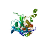
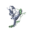
| Structure viewer | Molecule: MolmilJmol/JSmol |
|---|
- Downloads & links
-Download
| PDBx/mmCIF format | 1eg2.cif.gz | 73.6 KB | Display | PDBx/mmCIF format |
|---|---|---|---|---|
| PDB format | pdb1eg2.ent.gz | 54.1 KB | Display | PDB format |
| PDBx/mmJSON format | 1eg2.json.gz | Tree view | PDBx/mmJSON format | |
| Others |  Other downloads Other downloads |
-Validation report
| Arichive directory | https://data.pdbj.org/pub/pdb/validation_reports/eg/1eg2ftp://data.pdbj.org/pub/pdb/validation_reports/eg/1eg2 | HTTPS FTP |
|---|
-Related structure data
| Similar structure data | |
|---|---|
| Other databases |


-Links
 PDBj
PDBj- Assembly
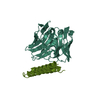
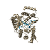
| Deposited unit | 
| ||||||||
|---|---|---|---|---|---|---|---|---|---|
| 1 |
| ||||||||
| 2 | 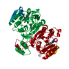
| ||||||||
| Unit cell |
|
-Components
| #1: Protein | Mass: 35702.191 Da / Num. of mol.: 1 Source method: isolated from a genetically manipulated source Source: (gene. exp.) Rhodobacter sphaeroides (bacteria) / Plasmid: PET28AReferences: UniProt: P14751, site-specific DNA-methyltransferase (adenine-specific) |
|---|---|
| #2: Chemical | ChemComp-MTA / 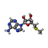  Mass: 297.334 Da / Num. of mol.: 1 / Source method: obtained synthetically / Formula: C11H15N5O3S Mass: 297.334 Da / Num. of mol.: 1 / Source method: obtained synthetically / Formula: C11H15N5O3S |
| #3: Water | ChemComp-HOH /  Mass: 18.015 Da / Num. of mol.: 227 / Source method: isolated from a natural source / Formula: H2O Mass: 18.015 Da / Num. of mol.: 227 / Source method: isolated from a natural source / Formula: H2O |
-Experimental details
-Experiment
| Experiment | Method: X-RAY DIFFRACTION / Number of used crystals: 2 |
|---|
- Sample preparation
| Crystal | Density Matthews: 2.16 Å3/Da / Density % sol: 43.06 % | ||||||||||||||||||||||||
|---|---|---|---|---|---|---|---|---|---|---|---|---|---|---|---|---|---|---|---|---|---|---|---|---|---|
| Crystal grow | Temperature: 291 K / Method: vapor diffusion, hanging drop / pH: 7.6 Details: 1.5 M Li2SO4, 0.1 M HEPES, 0.5% NaAzide, 10 mM EDTA, pH 7.6, VAPOR DIFFUSION, HANGING DROP, temperature 291K | ||||||||||||||||||||||||
| Crystal grow | *PLUS pH: 7.4 / Method: unknown | ||||||||||||||||||||||||
| Components of the solutions | *PLUS
|
-Data collection
| Diffraction |
| |||||||||||||||
|---|---|---|---|---|---|---|---|---|---|---|---|---|---|---|---|---|
| Diffraction source |
| |||||||||||||||
| Detector |
| |||||||||||||||
| Radiation | Protocol: SINGLE WAVELENGTH / Monochromatic (M) / Laue (L): M / Scattering type: x-ray | |||||||||||||||
| Radiation wavelength | Wavelength: 1 Å / Relative weight: 1 | |||||||||||||||
| Reflection | Resolution: 1.7→35.33 Å / Num. all: 188165 / Num. obs: 68339 / % possible obs: 94.2 % / Observed criterion σ(F): 0 / Observed criterion σ(I): 0 / Redundancy: 5.6 % / Biso Wilson estimate: 18.9 Å2 / Rmerge(I) obs: 0.035 / Net I/σ(I): 13.2 | |||||||||||||||
| Reflection shell | Resolution: 1.75→1.81 Å / Redundancy: 2.6 % / Rmerge(I) obs: 0.242 / Num. unique all: 2281 / % possible all: 93.9 | |||||||||||||||
| Reflection | *PLUS Highest resolution: 1.75 Å / Lowest resolution: 20 Å / Num. obs: 20741 / Num. measured all: 69339 | |||||||||||||||
| Reflection shell | *PLUS % possible obs: 93.9 % / Num. unique obs: 2281 / Num. measured obs: 2873 / Mean I/σ(I) obs: 4.3 |

- Processing
| Software |
| |||||||||||||||||||||||||
|---|---|---|---|---|---|---|---|---|---|---|---|---|---|---|---|---|---|---|---|---|---|---|---|---|---|---|
| Refinement | Resolution: 1.75→35.21 Å / σ(F): 0 / σ(I): 2 Stereochemistry target values: bond violations=0.05 angle violations=8.0 dihedral violations=60.0 improper violations=3.0 reported close contacts=2.5 lower resolution limit for coordinate error estimation=5.0
| |||||||||||||||||||||||||
| Refinement step | Cycle: LAST / Resolution: 1.75→35.21 Å
| |||||||||||||||||||||||||
| Refine LS restraints |
| |||||||||||||||||||||||||
| Software | *PLUS Name: CNS / Classification: refinement | |||||||||||||||||||||||||
| Refinement | *PLUS Rfactor Rfree: 0.24 / Rfactor Rwork: 0.21 | |||||||||||||||||||||||||
| Solvent computation | *PLUS | |||||||||||||||||||||||||
| Displacement parameters | *PLUS | |||||||||||||||||||||||||
| Refine LS restraints | *PLUS
| |||||||||||||||||||||||||
| LS refinement shell | *PLUS Highest resolution: 1.75 Å / Lowest resolution: 1.81 Å / Rfactor Rfree: 0.28 / Rfactor Rwork: 0.27 |
