 Movie
Movie Controller

[English] 日本語
 Yorodumi
- PDB-1ee9: CRYSTAL STRUCTURE OF THE NAD-DEPENDENT 5,10-METHYLENETETRAHYDROFO... -
+ Open data

- Basic information
| Entry | Database: PDB / ID: 1ee9 | ||||||
|---|---|---|---|---|---|---|---|
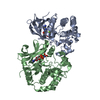
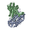
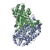
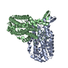
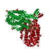
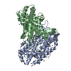
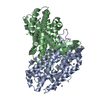
| Title | CRYSTAL STRUCTURE OF THE NAD-DEPENDENT 5,10-METHYLENETETRAHYDROFOLATE DEHYDROGENASE FROM SACCHAROMYCES CEREVISIAE COMPLEXED WITH NAD | ||||||
 Components Components | 5,10-METHYLENETETRAHYDROFOLATE DEHYDROGENASE | ||||||
 Keywords Keywords | OXIDOREDUCTASE / NUCLEOTIDE-BINDING DOMAIN / PROTEIN-NAD COMPLEX / MONOFUNCTIONAL / DEHYDROGENASE / FOLATE | ||||||
| Function / homology |  Function and homology information Function and homology informationmethylenetetrahydrofolate dehydrogenase (NAD+) / methylenetetrahydrofolate dehydrogenase (NAD+) activity / folic acid-containing compound biosynthetic process / purine nucleobase biosynthetic process / methylenetetrahydrofolate dehydrogenase (NADP+) activity / purine nucleotide biosynthetic process / one-carbon metabolic process / nucleus / cytosol Similarity search - Function | ||||||
| Biological species |  | ||||||
| Method |  X-RAY DIFFRACTION / DIFFERENCE FOURIER / Resolution: 3 Å X-RAY DIFFRACTION / DIFFERENCE FOURIER / Resolution: 3 Å | ||||||
 Authors Authors | Monzingo, A.F. / Breksa, A. / Ernst, S. / Appling, D.R. / Robertus, J.D. | ||||||
 Citation Citation | Journal: Protein Sci. / Year: 2000 Title: The X-ray structure of the NAD-dependent 5,10-methylenetetrahydrofolate dehydrogenase from Saccharomyces cerevisiae. Authors: Monzingo, A.F. / Breksa, A. / Ernst, S. / Appling, D.R. / Robertus, J.D. #1: Journal: Proteins / Year: 1996Title: Crystallization of the NAD-dependent 5,10-methylenetetrahydrofolate Dehydrogenase from Saccharomyces cerevisiae Authors: Monzingo, A.F. / West, M.G. / Schelp, E. / Appling, D.R. / Robertus, J.D. | ||||||
| History |
|
- Structure visualization
| Structure viewer | Molecule: MolmilJmol/JSmol |
|---|
- Downloads & links
-Download
| PDBx/mmCIF format | 1ee9.cif.gz | 69.7 KB | Display | PDBx/mmCIF format |
|---|---|---|---|---|
| PDB format | pdb1ee9.ent.gz | 52.5 KB | Display | PDB format |
| PDBx/mmJSON format | 1ee9.json.gz | Tree view | PDBx/mmJSON format | |
| Others |  Other downloads Other downloads |
-Validation report
| Arichive directory | https://data.pdbj.org/pub/pdb/validation_reports/ee/1ee9ftp://data.pdbj.org/pub/pdb/validation_reports/ee/1ee9 | HTTPS FTP |
|---|
-Related structure data


-Links
 PDBj
PDBj- Assembly
| Deposited unit | 
| ||||||||
|---|---|---|---|---|---|---|---|---|---|
| 1 | 
| ||||||||
| Unit cell |
|
-Components
| #1: Protein | Mass: 36284.562 Da / Num. of mol.: 1 Source method: isolated from a genetically manipulated source Source: (gene. exp.) Production host:  References: UniProt: Q02046, methylenetetrahydrofolate dehydrogenase (NAD+) |
|---|---|
| #2: Chemical | ChemComp-NAD /   Mass: 663.425 Da / Num. of mol.: 1 / Source method: obtained synthetically / Formula: C21H27N7O14P2 / Comment: NAD*YM Mass: 663.425 Da / Num. of mol.: 1 / Source method: obtained synthetically / Formula: C21H27N7O14P2 / Comment: NAD*YM |
| #3: Water | ChemComp-HOH /  Mass: 18.015 Da / Num. of mol.: 6 / Source method: isolated from a natural source / Formula: H2O Mass: 18.015 Da / Num. of mol.: 6 / Source method: isolated from a natural source / Formula: H2O |
-Experimental details
-Experiment
| Experiment | Method: X-RAY DIFFRACTION / Number of used crystals: 1 |
|---|
- Sample preparation
| Crystal | Density Matthews: 3.01 Å3/Da / Density % sol: 59.2 % | |||||||||||||||
|---|---|---|---|---|---|---|---|---|---|---|---|---|---|---|---|---|
| Crystal grow | Temperature: 277 K / Method: vapor diffusion, sitting drop / pH: 8.2 Details: PEG 8000, Tris-HCl, pH 8.2, VAPOR DIFFUSION, SITTING DROP, temperature 277K | |||||||||||||||
| Crystal grow | *PLUS Temperature: 4 ℃ / Method: vapor diffusion, hanging dropDetails: Monzingo, A.F., (1996) Proteins: Struct., Funct., Genet., 26, 481. | |||||||||||||||
| Components of the solutions | *PLUS
|
-Data collection
| Diffraction | Mean temperature: 100 K |
|---|---|
| Diffraction source | Source: ROTATING ANODE / Type: RIGAKU RU200 / Wavelength: 1.5418 |
| Detector | Type: RIGAKU RAXIS IV / Detector: IMAGE PLATE / Date: Apr 9, 1999 |
| Radiation | Protocol: SINGLE WAVELENGTH / Monochromatic (M) / Laue (L): M / Scattering type: x-ray |
| Radiation wavelength | Wavelength: 1.5418 Å / Relative weight: 1 |
| Reflection | Resolution: 2.9→30 Å / Num. all: 8474 / Num. obs: 8474 / % possible obs: 81.3 % / Observed criterion σ(I): 0 / Redundancy: 3.3 % / Rmerge(I) obs: 0.077 / Net I/σ(I): 11.3 |
| Reflection shell | Resolution: 2.9→3 Å / Redundancy: 3 % / Rmerge(I) obs: 0.578 / Num. unique all: 726 / % possible all: 73 |
| Reflection shell | *PLUS Mean I/σ(I) obs: 2.1 |
- Processing
| Software |
| |||||||||||||||||||||||||
|---|---|---|---|---|---|---|---|---|---|---|---|---|---|---|---|---|---|---|---|---|---|---|---|---|---|---|
| Refinement | Method to determine structure: DIFFERENCE FOURIER / Resolution: 3→20 Å / σ(F): 0 / Stereochemistry target values: Engh & Huber
| |||||||||||||||||||||||||
| Refinement step | Cycle: LAST / Resolution: 3→20 Å
| |||||||||||||||||||||||||
| Refine LS restraints |
| |||||||||||||||||||||||||
| Software | *PLUS Name: X-PLOR / Version: 3.851 / Classification: refinement | |||||||||||||||||||||||||
| Refinement | *PLUS Rfactor all: 0.25 / Rfactor obs: 0.25 / Rfactor Rwork: 0.25 | |||||||||||||||||||||||||
| Solvent computation | *PLUS | |||||||||||||||||||||||||
| Displacement parameters | *PLUS |
