 Movie
Movie Controller

[English] 日本語
 Yorodumi
- PDB-1e7u: Structure determinants of phosphoinositide 3-kinase inhibition by... -
+ Open data

- Basic information
| Entry | Database: PDB / ID: 1e7u | ||||||
|---|---|---|---|---|---|---|---|
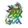
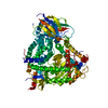
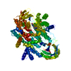
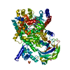
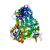
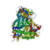
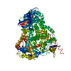
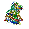
| Title | Structure determinants of phosphoinositide 3-kinase inhibition by wortmannin, LY294002, quercetin, myricetin and staurosporine | ||||||
 Components Components | PHOSPHATIDYLINOSITOL 3-KINASE CATALYTIC SUBUNIT | ||||||
 Keywords Keywords | PHOSPHOINOSITIDE 3-KINASE GAMMA / SECONDARY MESSENGER GENERATION / PI3K / PI 3K / WORTMANNIN | ||||||
| Function / homology |  Function and homology information Function and homology informationphosphatidylinositol-4-phosphate 3-kinase / phosphatidylinositol 3-kinase complex, class IB / sphingosine-1-phosphate receptor signaling pathway / 1-phosphatidylinositol-4-phosphate 3-kinase activity / phosphatidylinositol 3-kinase complex, class IA / phosphatidylinositol-4,5-bisphosphate 3-kinase / 1-phosphatidylinositol-4,5-bisphosphate 3-kinase activity / phosphatidylinositol 3-kinase / phosphatidylinositol-3-phosphate biosynthetic process / 1-phosphatidylinositol-3-kinase activity ...phosphatidylinositol-4-phosphate 3-kinase / phosphatidylinositol 3-kinase complex, class IB / sphingosine-1-phosphate receptor signaling pathway / 1-phosphatidylinositol-4-phosphate 3-kinase activity / phosphatidylinositol 3-kinase complex, class IA / phosphatidylinositol-4,5-bisphosphate 3-kinase / 1-phosphatidylinositol-4,5-bisphosphate 3-kinase activity / phosphatidylinositol 3-kinase / phosphatidylinositol-3-phosphate biosynthetic process / 1-phosphatidylinositol-3-kinase activity / phosphatidylinositol phosphate biosynthetic process / phosphatidylinositol-mediated signaling / positive regulation of Rac protein signal transduction / immune system process / positive regulation of endothelial cell migration / phosphatidylinositol 3-kinase/protein kinase B signal transduction / chemotaxis / endocytosis / cell migration / angiogenesis / phospholipase C-activating G protein-coupled receptor signaling pathway / non-specific serine/threonine protein kinase / inflammatory response / protein serine kinase activity / protein serine/threonine kinase activity / ATP binding / plasma membrane / cytosol / cytoplasm Similarity search - Function | ||||||
| Biological species |  | ||||||
| Method |  X-RAY DIFFRACTION / SYNCHROTRON / MOLECULAR REPLACEMENT / Resolution: 2 Å X-RAY DIFFRACTION / SYNCHROTRON / MOLECULAR REPLACEMENT / Resolution: 2 Å | ||||||
 Authors Authors | Walker, E.H. / Perisic, O. / Ried, C. / Stephens, L. / Williams, R.L. | ||||||
 Citation Citation | Journal: Mol.Cell / Year: 2000 Title: Structural Determinations of Phosphoinositide 3-Kinase Inhibition by Wortmannin, Ly294002, Quercetin, Myricetin and Staurosporine Authors: Walker, E.H. / Pacold, M.E. / Perisic, O. / Stephens, L. / Hawkins, P.T. / Whymann, M.P. / Williams, R.L. #1: Journal: Nature / Year: 1999 Title: Structural Insights Into Phosphoinositide 3-Kinase Catalysis and Signalling Authors: Walker, E.H. / Perisic, O. / Ried, C. / Stephens, L. / Williams, R.L. | ||||||
| History |
|
- Structure visualization
| Structure viewer | Molecule: MolmilJmol/JSmol |
|---|
- Downloads & links
-Download
| PDBx/mmCIF format | 1e7u.cif.gz | 192.6 KB | Display | PDBx/mmCIF format |
|---|---|---|---|---|
| PDB format | pdb1e7u.ent.gz | 150.4 KB | Display | PDB format |
| PDBx/mmJSON format | 1e7u.json.gz | Tree view | PDBx/mmJSON format | |
| Others |  Other downloads Other downloads |
-Validation report
| Arichive directory | https://data.pdbj.org/pub/pdb/validation_reports/e7/1e7uftp://data.pdbj.org/pub/pdb/validation_reports/e7/1e7u | HTTPS FTP |
|---|
-Related structure data
| Related structure data | 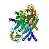 1e7vC 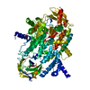 1e8wC  1e8xC 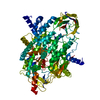 1e8yC 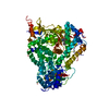 1e8zC  1e90C  1qmm S: Starting model for refinement C: citing same article ( |
|---|---|
| Similar structure data |


-Links
 PDBj
PDBj


- Assembly
| Deposited unit | 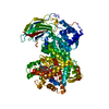
| ||||||||
|---|---|---|---|---|---|---|---|---|---|
| 1 |
| ||||||||
| Unit cell |
|
-Components
| #1: Protein | Mass: 109952.555 Da / Num. of mol.: 1 / Fragment: PI3-KINASE P110 SUBUNIT GAMMA / Mutation: YES Source method: isolated from a genetically manipulated source Details: WORTMANNIN / Source: (gene. exp.)   SPODOPTERA FRUGIPERDA (fall armyworm) / Strain (production host): SF9 / References: UniProt: O02697, phosphatidylinositol 3-kinase SPODOPTERA FRUGIPERDA (fall armyworm) / Strain (production host): SF9 / References: UniProt: O02697, phosphatidylinositol 3-kinase |
|---|---|
| #2: Chemical | ChemComp-KWT / (  Mass: 428.432 Da / Num. of mol.: 1 / Source method: obtained synthetically / Formula: C23H24O8 / Comment: inhibitor*YM Mass: 428.432 Da / Num. of mol.: 1 / Source method: obtained synthetically / Formula: C23H24O8 / Comment: inhibitor*YM |
| #3: Water | ChemComp-HOH /  Mass: 18.015 Da / Num. of mol.: 164 / Source method: isolated from a natural source / Formula: H2O Mass: 18.015 Da / Num. of mol.: 164 / Source method: isolated from a natural source / Formula: H2O |
| Has protein modification | Y |
| Sequence details | FUTHER DATA COLLECTION AND REFINEMENT AFTER THE ORIGINAL DEPOSITION OF 1QMM SUGGESTED A REGISTER ...FUTHER DATA COLLECTION |
-Experimental details
-Experiment
| Experiment | Method: X-RAY DIFFRACTION / Number of used crystals: 1 |
|---|
- Sample preparation
| Crystal | Density Matthews: 2.796 Å3/Da / Density % sol: 56.96 % | ||||||||||||||||||||
|---|---|---|---|---|---|---|---|---|---|---|---|---|---|---|---|---|---|---|---|---|---|
| Crystal grow | pH: 7.25 / Details: 14% PEG 4000, 0.2 M LI2SO4, 0.1 M TRIS PH 7.25 | ||||||||||||||||||||
| Crystal grow | *PLUS Temperature: 17 ℃ / pH: 7.2 / Method: vapor diffusion, hanging drop / Details: used hair seeding | ||||||||||||||||||||
| Components of the solutions | *PLUS
|
-Data collection
| Diffraction | Mean temperature: 100 K |
|---|---|
| Diffraction source | Source: SYNCHROTRON / Site: ESRF  / Beamline: ID14-2 / Wavelength: 0.93 / Beamline: ID14-2 / Wavelength: 0.93 |
| Detector | Type: MARRESEARCH / Detector: CCD / Date: Sep 15, 1999 / Details: BENT MIRROR |
| Radiation | Monochromator: DIAMOND C(111) / Protocol: SINGLE WAVELENGTH / Monochromatic (M) / Laue (L): M / Scattering type: x-ray |
| Radiation wavelength | Wavelength: 0.93 Å / Relative weight: 1 |
| Reflection | Resolution: 2→72.38 Å / Num. obs: 68705 / % possible obs: 97.4 % / Observed criterion σ(I): 0 / Redundancy: 3.3 % / Biso Wilson estimate: 30.8 Å2 / Rmerge(I) obs: 0.083 / Net I/σ(I): 16.01 |
| Reflection shell | Resolution: 2→2.11 Å / Redundancy: 3 % / Mean I/σ(I) obs: 1.6 / Rsym value: 0.575 / % possible all: 94.2 |
| Reflection | *PLUS Num. measured all: 227525 |
| Reflection shell | *PLUS Rmerge(I) obs: 94.2 / Mean I/σ(I) obs: 1.1 |
- Processing
| Software |
| ||||||||||||||||||||||||||||||||||||||||||||||||||||||||||||||||||||||||||||||||
|---|---|---|---|---|---|---|---|---|---|---|---|---|---|---|---|---|---|---|---|---|---|---|---|---|---|---|---|---|---|---|---|---|---|---|---|---|---|---|---|---|---|---|---|---|---|---|---|---|---|---|---|---|---|---|---|---|---|---|---|---|---|---|---|---|---|---|---|---|---|---|---|---|---|---|---|---|---|---|---|---|---|
| Refinement | Method to determine structure: MOLECULAR REPLACEMENT Starting model: 1QMM 1qmm Resolution: 2→72.38 Å / Rfactor Rfree error: 0.006 / Data cutoff high absF: 2435589.98 / Isotropic thermal model: RESTRAINED / Cross valid method: THROUGHOUT / σ(F): 0
| ||||||||||||||||||||||||||||||||||||||||||||||||||||||||||||||||||||||||||||||||
| Solvent computation | Solvent model: FLAT MODEL / Bsol: 44.0509 Å2 / ksol: 0.341002 e/Å3 | ||||||||||||||||||||||||||||||||||||||||||||||||||||||||||||||||||||||||||||||||
| Displacement parameters | Biso mean: 49.9 Å2
| ||||||||||||||||||||||||||||||||||||||||||||||||||||||||||||||||||||||||||||||||
| Refine analyze |
| ||||||||||||||||||||||||||||||||||||||||||||||||||||||||||||||||||||||||||||||||
| Refinement step | Cycle: LAST / Resolution: 2→72.38 Å
| ||||||||||||||||||||||||||||||||||||||||||||||||||||||||||||||||||||||||||||||||
| Refine LS restraints |
| ||||||||||||||||||||||||||||||||||||||||||||||||||||||||||||||||||||||||||||||||
| LS refinement shell | Resolution: 2→2.13 Å / Rfactor Rfree error: 0.014 / Total num. of bins used: 6
| ||||||||||||||||||||||||||||||||||||||||||||||||||||||||||||||||||||||||||||||||
| Xplor file |
| ||||||||||||||||||||||||||||||||||||||||||||||||||||||||||||||||||||||||||||||||
| Software | *PLUS Name: CNS / Version: 1 / Classification: refinement | ||||||||||||||||||||||||||||||||||||||||||||||||||||||||||||||||||||||||||||||||
| Refine LS restraints | *PLUS
|
