 Movie
Movie Controller

[English] 日本語
 Yorodumi
- PDB-1e59: E.coli cofactor-dependent phosphoglycerate mutase complexed with ... -
+ Open data

- Basic information
| Entry | Database: PDB / ID: 1.0E+59 | ||||||
|---|---|---|---|---|---|---|---|
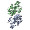
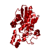
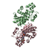
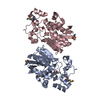
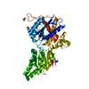
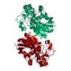
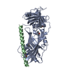
| Title | E.coli cofactor-dependent phosphoglycerate mutase complexed with vanadate | ||||||
 Components Components | PHOSPHOGLYCERATE MUTASE | ||||||
 Keywords Keywords | ISOMERASE / INHIBITOR / VANDATE / GLYCOLYSIS AND GLUCONEOGENESIS / PHOSPHOGLYCERATE MUTASE | ||||||
| Function / homology |  Function and homology information Function and homology information: / phosphoglycerate mutase (2,3-diphosphoglycerate-dependent) / phosphoglycerate mutase activity / guanosine tetraphosphate binding / canonical glycolysis / gluconeogenesis / protein homodimerization activity / ATP binding / cytoplasm / cytosol Similarity search - Function | ||||||
| Biological species |  | ||||||
| Method |  X-RAY DIFFRACTION / SYNCHROTRON / MOLECULAR REPLACEMENT / Resolution: 1.3 Å X-RAY DIFFRACTION / SYNCHROTRON / MOLECULAR REPLACEMENT / Resolution: 1.3 Å | ||||||
 Authors Authors | Bond, C.S. / Hunter, W.N. | ||||||
 Citation Citation | Journal: J.Mol.Biol. / Year: 2002 Title: Mechanistic Implications for Escherichia Coli Cofactor-Dependent Phosphoglycerate Mutase Based on the High-Resolution Crystal Structure of a Vanadate Complex. Authors: Bond, C.S. / White, M. / Hunter, W.N. | ||||||
| History |
|
- Structure visualization
| Structure viewer | Molecule: MolmilJmol/JSmol |
|---|
- Downloads & links
-Download
| PDBx/mmCIF format | 1e59.cif.gz | 122.8 KB | Display | PDBx/mmCIF format |
|---|---|---|---|---|
| PDB format | pdb1e59.ent.gz | 94.6 KB | Display | PDB format |
| PDBx/mmJSON format | 1e59.json.gz | Tree view | PDBx/mmJSON format | |
| Others |  Other downloads Other downloads |
-Validation report
| Arichive directory | https://data.pdbj.org/pub/pdb/validation_reports/e5/1e59ftp://data.pdbj.org/pub/pdb/validation_reports/e5/1e59 | HTTPS FTP |
|---|
-Related structure data
| Related structure data |  1e58S S: Starting model for refinement |
|---|---|
| Similar structure data |


-Links
 PDBj
PDBj


- Assembly
| Deposited unit | 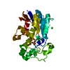
| ||||||||
|---|---|---|---|---|---|---|---|---|---|
| 1 | 
| ||||||||
| Unit cell |
|
-Components
| #1: Protein | Mass: 28465.195 Da / Num. of mol.: 1 Source method: isolated from a genetically manipulated source Source: (gene. exp.) References: UniProt: P31217, UniProt: P62707*PLUS, EC: 5.4.2.1 |
|---|---|
| #2: Chemical | ChemComp-VO3 /   Mass: 411.758 Da / Num. of mol.: 1 / Source method: obtained synthetically / Formula: O13V4 Mass: 411.758 Da / Num. of mol.: 1 / Source method: obtained synthetically / Formula: O13V4 |
| #3: Chemical | ChemComp-CL /   Mass: 35.453 Da / Num. of mol.: 1 / Source method: obtained synthetically / Formula: Cl Mass: 35.453 Da / Num. of mol.: 1 / Source method: obtained synthetically / Formula: Cl |
| #4: Water | ChemComp-HOH /  Mass: 18.015 Da / Num. of mol.: 264 / Source method: isolated from a natural source / Formula: H2O Mass: 18.015 Da / Num. of mol.: 264 / Source method: isolated from a natural source / Formula: H2O |
| Compound details | CATALYTIC ACTIVITY: 2-PHOSPHOGLYCERATE + 2,3-DIPHOSPHOGLYCERATE = 3-PHOSPHOGLYCERATE + 2,3- ...CATALYTIC ACTIVITY: 2-PHOSPHOGLY |
-Experimental details
-Experiment
| Experiment | Method: X-RAY DIFFRACTION / Number of used crystals: 1 |
|---|
- Sample preparation
| Crystal | Density Matthews: 2.21 Å3/Da / Density % sol: 51 % | ||||||||||||||||||||||||||||||||||||||||||||||||||||||||
|---|---|---|---|---|---|---|---|---|---|---|---|---|---|---|---|---|---|---|---|---|---|---|---|---|---|---|---|---|---|---|---|---|---|---|---|---|---|---|---|---|---|---|---|---|---|---|---|---|---|---|---|---|---|---|---|---|---|
| Crystal grow | pH: 8.5 Details: 100 MM TRIS-HCL (PH 8.0), 200 MM LI2SO4, 20% PEG 4000, 100 MM NAVO3 | ||||||||||||||||||||||||||||||||||||||||||||||||||||||||
| Crystal grow | *PLUS pH: 8 / Method: vapor diffusion, hanging drop | ||||||||||||||||||||||||||||||||||||||||||||||||||||||||
| Components of the solutions | *PLUS
|
-Data collection
| Diffraction | Mean temperature: 105 K |
|---|---|
| Diffraction source | Source: SYNCHROTRON / Site: SRS  / Beamline: PX9.6 / Wavelength: 0.89 / Beamline: PX9.6 / Wavelength: 0.89 |
| Detector | Date: Sep 15, 1999 |
| Radiation | Protocol: SINGLE WAVELENGTH / Monochromatic (M) / Laue (L): M / Scattering type: x-ray |
| Radiation wavelength | Wavelength: 0.89 Å / Relative weight: 1 |
| Reflection | Resolution: 1.3→30 Å / Num. obs: 68073 / % possible obs: 96.6 % / Redundancy: 4 % / Rsym value: 0.079 / Net I/σ(I): 17 |
| Reflection shell | Resolution: 1.3→1.31 Å / Redundancy: 3 % / Mean I/σ(I) obs: 2.1 / Rsym value: 0.401 / % possible all: 99.9 |
| Reflection | *PLUS Redundancy: 3.1 % / Rmerge(I) obs: 0.079 |
| Reflection shell | *PLUS % possible obs: 99.9 % / Redundancy: 3.1 % / Rmerge(I) obs: 0.401 |
- Processing
| Software |
| |||||||||||||||||||||||||||||||||
|---|---|---|---|---|---|---|---|---|---|---|---|---|---|---|---|---|---|---|---|---|---|---|---|---|---|---|---|---|---|---|---|---|---|---|
| Refinement | Method to determine structure: MOLECULAR REPLACEMENT Starting model: PDB ENTRY 1E58 Resolution: 1.3→30 Å / Num. parameters: 20185 / Num. restraintsaints: 24690 / Cross valid method: FREE R-VALUE / σ(F): 0 StereochEM target val spec case: VO3 VALUES EXTRACTED FROM CSDS Stereochemistry target values: ENGH AND HUBER
| |||||||||||||||||||||||||||||||||
| Solvent computation | Solvent model: MOEWS & KRETSINGER, J.MOL.BIOL.91(1973)201-2 | |||||||||||||||||||||||||||||||||
| Refine analyze | Num. disordered residues: 4 / Occupancy sum hydrogen: 0 / Occupancy sum non hydrogen: 2210.5 | |||||||||||||||||||||||||||||||||
| Refinement step | Cycle: LAST / Resolution: 1.3→30 Å
| |||||||||||||||||||||||||||||||||
| Refine LS restraints |
| |||||||||||||||||||||||||||||||||
| Software | *PLUS Name: SHELXL / Version: 97 / Classification: refinement | |||||||||||||||||||||||||||||||||
| Refinement | *PLUS Rfactor Rwork: 0.1552 | |||||||||||||||||||||||||||||||||
| Solvent computation | *PLUS | |||||||||||||||||||||||||||||||||
| Displacement parameters | *PLUS | |||||||||||||||||||||||||||||||||
| Refine LS restraints | *PLUS
|
