 Movie
Movie Controller


+ Open data

- Basic information
| Entry | Database: PDB / ID: 1e3b | ||||||
|---|---|---|---|---|---|---|---|
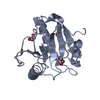
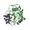
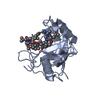
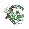
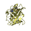
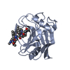
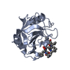
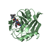
| Title | CYCLOPHILIN 3 FROM C.ELEGANS COMPLEXED WITH AUP(ET)3 | ||||||
 Components Components | CYCLOPHILIN 3 | ||||||
 Keywords Keywords | ISOMERASE / AUPET3 ADDUCT CYCLOPHILIN ANTIARTHRITIC GOLD | ||||||
| Function / homology |  Function and homology information Function and homology informationcyclosporin A binding / peptidylprolyl isomerase / peptidyl-prolyl cis-trans isomerase activity / protein folding / cytoplasm Similarity search - Function | ||||||
| Biological species |  | ||||||
| Method |  X-RAY DIFFRACTION / SYNCHROTRON / MOLECULAR REPLACEMENT / Resolution: 1.85 Å X-RAY DIFFRACTION / SYNCHROTRON / MOLECULAR REPLACEMENT / Resolution: 1.85 Å | ||||||
 Authors Authors | Zou, J. / Taylor, P. / Dornan, J. / Robinson, S.P. / Walkinshaw, M.D. / Sadler, P.J. | ||||||
 Citation Citation | Journal: Angew.Chem.Int.Ed.Engl. / Year: 2000 Title: First Crystal Structure of a Medicinally Relevant Gold Protein Complex:Unexpected Binding of [Au(Pet (3))](+) to Histidine Authors: Zou, J. / Taylor, P. / Dornan, J. / Robinson, S.P. / Walkinshaw, M.D. / Sadler, P.J. | ||||||
| History |
|
- Structure visualization
| Structure viewer | Molecule: MolmilJmol/JSmol |
|---|
- Downloads & links
-Download
| PDBx/mmCIF format | 1e3b.cif.gz | 69.8 KB | Display | PDBx/mmCIF format |
|---|---|---|---|---|
| PDB format | pdb1e3b.ent.gz | 49.4 KB | Display | PDB format |
| PDBx/mmJSON format | 1e3b.json.gz | Tree view | PDBx/mmJSON format | |
| Others |  Other downloads Other downloads |
-Validation report
| Arichive directory | https://data.pdbj.org/pub/pdb/validation_reports/e3/1e3bftp://data.pdbj.org/pub/pdb/validation_reports/e3/1e3b | HTTPS FTP |
|---|
-Related structure data
| Similar structure data |
|---|


-Links
 PDBj
PDBj


- Assembly
| Deposited unit | 
| ||||||||
|---|---|---|---|---|---|---|---|---|---|
| 1 |
| ||||||||
| Unit cell |
|
-Components
| #1: Protein | Mass: 18576.182 Da / Num. of mol.: 1 Source method: isolated from a genetically manipulated source Source: (gene. exp.)  |
|---|---|
| #2: Chemical | ChemComp-AU /   Mass: 196.967 Da / Num. of mol.: 1 / Source method: obtained synthetically / Formula: Au Mass: 196.967 Da / Num. of mol.: 1 / Source method: obtained synthetically / Formula: Au |
| #3: Chemical | ChemComp-3EP /   Mass: 118.157 Da / Num. of mol.: 1 / Source method: obtained synthetically / Formula: C6H15P Mass: 118.157 Da / Num. of mol.: 1 / Source method: obtained synthetically / Formula: C6H15P |
| #4: Water | ChemComp-HOH /  Mass: 18.015 Da / Num. of mol.: 217 / Source method: isolated from a natural source / Formula: H2O Mass: 18.015 Da / Num. of mol.: 217 / Source method: isolated from a natural source / Formula: H2O |
-Experimental details
-Experiment
| Experiment | Method: X-RAY DIFFRACTION / Number of used crystals: 1 |
|---|
- Sample preparation
| Crystal | Density Matthews: 2.63 Å3/Da / Density % sol: 60.29 % | ||||||||||||||||||||||||||||||
|---|---|---|---|---|---|---|---|---|---|---|---|---|---|---|---|---|---|---|---|---|---|---|---|---|---|---|---|---|---|---|---|
| Crystal grow | Method: vapor diffusion, hanging drop / pH: 5.6 Details: HANGING DROP PH5.6 15.5% W/V MPEG 5000, 50 MM SODIUM CITRATE. WELL SOLUTION: 31% MPEG 5000, 100MM SODIUM CITRATE, pH 5.60 | ||||||||||||||||||||||||||||||
| Crystal grow | *PLUS Temperature: 291 K / Method: vapor diffusion, hanging drop | ||||||||||||||||||||||||||||||
| Components of the solutions | *PLUS
|
-Data collection
| Diffraction | Mean temperature: 100 K |
|---|---|
| Diffraction source | Source: SYNCHROTRON / Site: SRS  / Beamline: PX9.5 / Wavelength: 1.3 / Beamline: PX9.5 / Wavelength: 1.3 |
| Detector | Type: MARRESEARCH / Detector: CCD / Date: Sep 17, 1999 / Details: PT COATED TORROIDAL MIRRO |
| Radiation | Monochromator: SI(111) / Protocol: SINGLE WAVELENGTH / Monochromatic (M) / Laue (L): M / Scattering type: x-ray |
| Radiation wavelength | Wavelength: 1.3 Å / Relative weight: 1 |
| Reflection | Resolution: 1.85→35 Å / Num. obs: 20154 / % possible obs: 98.3 % / Redundancy: 4.12 % / Rmerge(I) obs: 0.068 / Net I/σ(I): 24.03 |
| Reflection shell | Resolution: 1.85→1.88 Å / Redundancy: 4.3 % / Rmerge(I) obs: 0.253 / Mean I/σ(I) obs: 9.92 / % possible all: 1 |
| Reflection | *PLUS Num. measured all: 185907 |
| Reflection shell | *PLUS % possible obs: 99.7 % |
- Processing
| Software |
| |||||||||||||||||||||||||||||||||
|---|---|---|---|---|---|---|---|---|---|---|---|---|---|---|---|---|---|---|---|---|---|---|---|---|---|---|---|---|---|---|---|---|---|---|
| Refinement | Method to determine structure: MOLECULAR REPLACEMENT Starting model: CYCLOPHILIN 3 NATIVE Resolution: 1.85→31 Å / Num. parameters: 6117 / Num. restraintsaints: 5315 / Cross valid method: FREE R-VALUE / σ(F): 0 StereochEM target val spec case: AU PET3 PARAMETERS CALCULATED FROM SMALL MOLECULE DATABASE (CCDC) Stereochemistry target values: ENGH AND HUBER Details: ANISOTROPIC REFINEMENT REDUCED FREE R (NO CUTOFF) BY 0.03
| |||||||||||||||||||||||||||||||||
| Solvent computation | Solvent model: MOEWS & KRETSINGER, J.MOL.BIOL.91(1973)201-2 | |||||||||||||||||||||||||||||||||
| Refine analyze | Num. disordered residues: 0 / Occupancy sum hydrogen: 0 / Occupancy sum non hydrogen: 1526 | |||||||||||||||||||||||||||||||||
| Refinement step | Cycle: LAST / Resolution: 1.85→31 Å
| |||||||||||||||||||||||||||||||||
| Refine LS restraints |
| |||||||||||||||||||||||||||||||||
| Software | *PLUS Name: SHELXL-97 / Classification: refinement | |||||||||||||||||||||||||||||||||
| Refine LS restraints | *PLUS Type: s_chiral_restr / Dev ideal: 0.036 |
