 Movie
Movie Controller

[English] 日本語
 Yorodumi
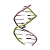
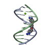
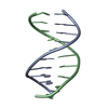
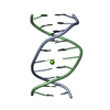
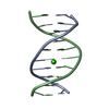
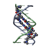
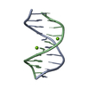
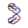
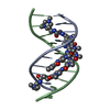
- PDB-1dvl: CRYSTAL STRUCTURE OF THE 1:1 NETROPSIN-DECAMER D(CCIICICCII)2 COM... -
+ Open data

- Basic information
| Entry | Database: PDB / ID: 1dvl | ||||||||||||||||||
|---|---|---|---|---|---|---|---|---|---|---|---|---|---|---|---|---|---|---|---|
| Title | CRYSTAL STRUCTURE OF THE 1:1 NETROPSIN-DECAMER D(CCIICICCII)2 COMPLEX WITH ONLY ONE DRUG BOUND AT ONE END | ||||||||||||||||||
 Components Components | 5'-D(* Keywords KeywordsDNA / DRUG BINDING | Function / homology | NETROPSIN / DNA |  Function and homology information Function and homology informationMethod |  X-RAY DIFFRACTION / MOLECULAR REPLACEMENT / Resolution: 2.4 Å X-RAY DIFFRACTION / MOLECULAR REPLACEMENT / Resolution: 2.4 Å  Authors AuthorsShi, K. / Mitra, S.N. / Sundaralingam, M. |  CitationJournal: Acta Crystallogr.,Sect.D / Year: 2002 CitationJournal: Acta Crystallogr.,Sect.D / Year: 2002Title: Structure of the 1:1 netropsin-decamer d(CCIICICCII)2 complex with a single bound netropsin. Authors: Shi, K. / Mitra, S.N. / Sundaralingam, M. History |
|
- Structure visualization
| Structure viewer | Molecule: MolmilJmol/JSmol |
|---|
- Downloads & links
-Download
| PDBx/mmCIF format | 1dvl.cif.gz | 17.8 KB | Display | PDBx/mmCIF format |
|---|---|---|---|---|
| PDB format | pdb1dvl.ent.gz | 12.8 KB | Display | PDB format |
| PDBx/mmJSON format | 1dvl.json.gz | Tree view | PDBx/mmJSON format | |
| Others |  Other downloads Other downloads |
-Validation report
| Arichive directory | https://data.pdbj.org/pub/pdb/validation_reports/dv/1dvlftp://data.pdbj.org/pub/pdb/validation_reports/dv/1dvl | HTTPS FTP |
|---|
-Related structure data
| Similar structure data |
|---|
-Links
 PDBj
PDBj

- Assembly
| Deposited unit | 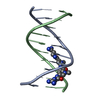
| ||||||||||
|---|---|---|---|---|---|---|---|---|---|---|---|
| 1 |
| ||||||||||
| Unit cell |
|
-Components
| #1: DNA chain | Mass: 2971.911 Da / Num. of mol.: 2 / Source method: obtained synthetically #2: Chemical | ChemComp-NT / |   Mass: 430.464 Da / Num. of mol.: 1 / Source method: obtained synthetically / Formula: C18H26N10O3 / Comment: antibiotic*YM Mass: 430.464 Da / Num. of mol.: 1 / Source method: obtained synthetically / Formula: C18H26N10O3 / Comment: antibiotic*YM#3: Water | ChemComp-HOH / |  Mass: 18.015 Da / Num. of mol.: 31 / Source method: isolated from a natural source / Formula: H2O Mass: 18.015 Da / Num. of mol.: 31 / Source method: isolated from a natural source / Formula: H2O |
|---|
-Experimental details
-Experiment
| Experiment | Method: X-RAY DIFFRACTION / Number of used crystals: 1 |
|---|
- Sample preparation
| Crystal | Density Matthews: 2.08 Å3/Da / Density % sol: 40.81 % | |||||||||||||||||||||||||||||||||||||||||||||||||
|---|---|---|---|---|---|---|---|---|---|---|---|---|---|---|---|---|---|---|---|---|---|---|---|---|---|---|---|---|---|---|---|---|---|---|---|---|---|---|---|---|---|---|---|---|---|---|---|---|---|---|
| Crystal grow | Temperature: 293 K / Method: vapor diffusion, hanging drop / pH: 6.5 Details: 4 MM DNA (SINGLE STRAND), 0.008M MGCL2, 0.04 M NA CACODYLATE (PH 6.5), 2MM NETROPSIN, VAPOR DIFFUSION, HANGING DROP at 293K | |||||||||||||||||||||||||||||||||||||||||||||||||
| Components of the solutions |
| |||||||||||||||||||||||||||||||||||||||||||||||||
| Crystal grow | *PLUS | |||||||||||||||||||||||||||||||||||||||||||||||||
| Components of the solutions | *PLUS
|
-Data collection
| Diffraction | Mean temperature: 293 K |
|---|---|
| Diffraction source | Source: ROTATING ANODE / Type: RIGAKU / Wavelength: 1.5418 |
| Detector | Type: RIGAKU RAXIS IIC / Detector: IMAGE PLATE |
| Radiation | Monochromator: GRAPHITE / Protocol: SINGLE WAVELENGTH / Monochromatic (M) / Laue (L): M / Scattering type: x-ray |
| Radiation wavelength | Wavelength: 1.5418 Å / Relative weight: 1 |
| Reflection | Highest resolution: 2.4 Å / Num. obs: 1795 / Observed criterion σ(I): 1 / Rmerge(I) obs: 0.037 |
| Reflection shell | Resolution: 2.4→2.5 Å |
| Reflection | *PLUS Highest resolution: 2.4 Å / Rmerge(I) obs: 0.037 |
| Reflection shell | *PLUS Highest resolution: 2.4 Å / Lowest resolution: 2.5 Å / % possible obs: 62.7 % |
- Processing
| Software |
| ||||||||||||||||||||||||||||||||||||||||||||||||||||||||||||
|---|---|---|---|---|---|---|---|---|---|---|---|---|---|---|---|---|---|---|---|---|---|---|---|---|---|---|---|---|---|---|---|---|---|---|---|---|---|---|---|---|---|---|---|---|---|---|---|---|---|---|---|---|---|---|---|---|---|---|---|---|---|
| Refinement | Method to determine structure: MOLECULAR REPLACEMENT / Resolution: 2.4→8 Å / Cross valid method: THROUGHOUT / σ(F): 2
| ||||||||||||||||||||||||||||||||||||||||||||||||||||||||||||
| Refinement step | Cycle: LAST / Resolution: 2.4→8 Å
| ||||||||||||||||||||||||||||||||||||||||||||||||||||||||||||
| Refine LS restraints |
| ||||||||||||||||||||||||||||||||||||||||||||||||||||||||||||
| Refinement | *PLUS % reflection Rfree: 10 % / Rfactor obs: 0.196 / Rfactor Rfree: 0.245 / Rfactor Rwork: 0.196 | ||||||||||||||||||||||||||||||||||||||||||||||||||||||||||||
| Solvent computation | *PLUS | ||||||||||||||||||||||||||||||||||||||||||||||||||||||||||||
| Displacement parameters | *PLUS | ||||||||||||||||||||||||||||||||||||||||||||||||||||||||||||
| Refine LS restraints | *PLUS
|
