 Movie
Movie Controller

[English] 日本語
 Yorodumi
- PDB-1dur: Replacement for 1FDX 2(4FE4S) ferredoxin from (NOW) Peptostreptoc... -
+ Open data

- Basic information
| Entry | Database: PDB / ID: 1dur | |||||||||
|---|---|---|---|---|---|---|---|---|---|---|
| Title | Replacement for 1FDX 2(4FE4S) ferredoxin from (NOW) Peptostreptococcus asaccharolyticus | |||||||||
 Components Components | 2[4FE-4S] FERREDOXIN | |||||||||
 Keywords Keywords | ELECTRON TRANSPORT / TWO 4Fe-4S clusters | |||||||||
| Function / homology |  Function and homology information Function and homology informationAlpha-Beta Plaits - #20 / 4Fe-4S binding domain / : / 4Fe-4S ferredoxin, iron-sulphur binding, conserved site / 4Fe-4S ferredoxin-type iron-sulfur binding region signature. / 4Fe-4S ferredoxin-type iron-sulfur binding domain profile. / 4Fe-4S ferredoxin-type, iron-sulphur binding domain / Alpha-Beta Plaits / 2-Layer Sandwich / Alpha Beta Similarity search - Domain/homology | |||||||||
| Biological species |  Peptoniphilus asaccharolyticus (bacteria) Peptoniphilus asaccharolyticus (bacteria) | |||||||||
| Method |  X-RAY DIFFRACTION / Resolution: 2 Å X-RAY DIFFRACTION / Resolution: 2 Å | |||||||||
 Authors Authors | Adman, E.T. / Sieker, L.C. | |||||||||
 Citation Citation | #1: Journal: J.Am.Chem.Soc. / Year: 1991Title: The Environment of Fe4S4 Clusters in Ferredoxins and High-Potential Iron Proteins. New Information from X-ray Crystallography and Resonance Raman Spectroscopy Authors: Backes, G. / Mino, Y. / Loehr, T.M. / Meyer, T.E. / Cusanovich, M. / Sweeney, W.V. / Adman, E.T. / Sanders-Loehr, J. #2: Journal: J.Biol.Chem. / Year: 1976Title: Structure of Peptococcus aerogenes Ferredoxin Refinement at 2 A Resolution Authors: Adman, E.T. / Sieker, L.C. / Jensen, L.H. #3: Journal: J.Biol.Chem. / Year: 1973Title: The Structure of a Bacterial Ferredoxin Authors: Adman, E.T. / Sieker, L.C. / Jensen, L.H. | |||||||||
| History |
|
- Structure visualization
| Structure viewer | Molecule: MolmilJmol/JSmol |
|---|
- Downloads & links
-Download
| PDBx/mmCIF format | 1dur.cif.gz | 24.7 KB | Display | PDBx/mmCIF format |
|---|---|---|---|---|
| PDB format | pdb1dur.ent.gz | 15 KB | Display | PDB format |
| PDBx/mmJSON format | 1dur.json.gz | Tree view | PDBx/mmJSON format | |
| Others |  Other downloads Other downloads |
-Validation report
| Arichive directory | https://data.pdbj.org/pub/pdb/validation_reports/du/1durftp://data.pdbj.org/pub/pdb/validation_reports/du/1dur | HTTPS FTP |
|---|
-Related structure data
| Similar structure data |
|---|
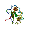



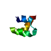

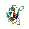

-Links
 PDBj
PDBj


- Assembly
| Deposited unit | 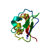
| ||||||||
|---|---|---|---|---|---|---|---|---|---|
| 1 |
| ||||||||
| Unit cell |
|
-Components
| #1: Protein | Mass: 5557.229 Da / Num. of mol.: 1 / Source method: isolated from a natural source / Details: FORMERLY CALLED PEPTOCOCCUS AEROGENES / Source: (natural) Peptoniphilus asaccharolyticus (bacteria) / References: UniProt: P00193 | ||||
|---|---|---|---|---|---|
| #2: Chemical |  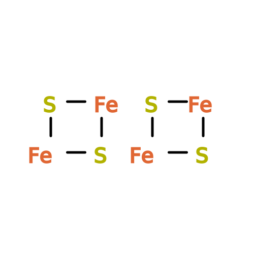 Mass: 351.640 Da / Num. of mol.: 2 / Source method: obtained synthetically / Formula: Fe4S4 Mass: 351.640 Da / Num. of mol.: 2 / Source method: obtained synthetically / Formula: Fe4S4#3: Water | ChemComp-HOH / |  Mass: 18.015 Da / Num. of mol.: 93 / Source method: isolated from a natural source / Formula: H2O Mass: 18.015 Da / Num. of mol.: 93 / Source method: isolated from a natural source / Formula: H2OHas protein modification | Y | |
-Experimental details
-Experiment
| Experiment | Method: X-RAY DIFFRACTION / Number of used crystals: 2 |
|---|
- Sample preparation
| Crystal | Density Matthews: 2.04 Å3/Da / Density % sol: 39.67 % |
|---|---|
| Crystal grow | Temperature: 277 K / Method: evaporation / pH: 7.5 Details: 3.3-3.5M ammonium sulfate, tris-chloride buffer, pH 7.5, EVAPORATION, temperature 277.0K |
-Data collection
| Diffraction | Mean temperature: 298 K |
|---|---|
| Diffraction source | Source: SEALED TUBE / Type: OTHER / Wavelength: 1.5418 |
| Detector | Type: PICKER / Detector: DIFFRACTOMETER / Date: Jan 1, 1972 |
| Radiation | Protocol: SINGLE WAVELENGTH / Monochromatic (M) / Laue (L): M / Scattering type: x-ray |
| Radiation wavelength | Wavelength: 1.5418 Å / Relative weight: 1 |
| Reflection | Resolution: 2→10 Å / Num. all: 3220 / Num. obs: 3220 / % possible obs: 100 % / Observed criterion σ(F): 0 / Observed criterion σ(I): 0 / Rmerge(I) obs: 0.05 |
- Processing
| Software |
| ||||||||||||
|---|---|---|---|---|---|---|---|---|---|---|---|---|---|
| Refinement | Resolution: 2→10 Å / σ(F): 0 / σ(I): 1 / Stereochemistry target values: Engh and Huber Details: Used Xplor positional and individual B refinement. Refinement with "modern" tools (PROLSQ, then XPLOR) revealed a new cysteine in position 22, subsequently verified by partial sequencing. ...Details: Used Xplor positional and individual B refinement. Refinement with "modern" tools (PROLSQ, then XPLOR) revealed a new cysteine in position 22, subsequently verified by partial sequencing. See Citation 1. The close contacts listed in remark 500 are waters that are regarded as alternative sites, not all occupied simultaneously.
| ||||||||||||
| Refinement step | Cycle: LAST / Resolution: 2→10 Å
|
