 Movie
Movie Controller

[English] 日本語
 Yorodumi
- PDB-1dtl: CRYSTAL STRUCTURE OF CALCIUM-SATURATED (3CA2+) CARDIAC TROPONIN C... -
+ Open data

- Basic information
| Entry | Database: PDB / ID: 1dtl | ||||||
|---|---|---|---|---|---|---|---|
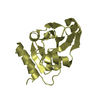
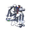
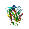
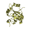
| Title | CRYSTAL STRUCTURE OF CALCIUM-SATURATED (3CA2+) CARDIAC TROPONIN C COMPLEXED WITH THE CALCIUM SENSITIZER BEPRIDIL AT 2.15 A RESOLUTION | ||||||
 Components Components | CARDIAC TROPONIN C | ||||||
 Keywords Keywords | STRUCTURAL PROTEIN / helix-turn-helix | ||||||
| Function / homology |  Function and homology information Function and homology informationcardiac Troponin complex / Striated Muscle Contraction / troponin I binding / skeletal muscle contraction / cardiac muscle contraction / calcium-dependent protein binding / calcium ion binding Similarity search - Function | ||||||
| Biological species |  | ||||||
| Method |  X-RAY DIFFRACTION / SYNCHROTRON / MOLECULAR REPLACEMENT / Resolution: 2.15 Å X-RAY DIFFRACTION / SYNCHROTRON / MOLECULAR REPLACEMENT / Resolution: 2.15 Å | ||||||
 Authors Authors | Li, Y. / Love, M.L. / Putkey, J.A. / Cohen, C. | ||||||
 Citation Citation | Journal: Proc.Natl.Acad.Sci.USA / Year: 2000 Title: Bepridil opens the regulatory N-terminal lobe of cardiac troponin C. Authors: Li, Y. / Love, M.L. / Putkey, J.A. / Cohen, C. | ||||||
| History |
|
- Structure visualization
| Structure viewer | Molecule: MolmilJmol/JSmol |
|---|
- Downloads & links
-Download
| PDBx/mmCIF format | 1dtl.cif.gz | 49.6 KB | Display | PDBx/mmCIF format |
|---|---|---|---|---|
| PDB format | pdb1dtl.ent.gz | 33.6 KB | Display | PDB format |
| PDBx/mmJSON format | 1dtl.json.gz | Tree view | PDBx/mmJSON format | |
| Others |  Other downloads Other downloads |
-Validation report
| Summary document | 1dtl_validation.pdf.gz | 574.1 KB | Display | wwPDB validaton report |
|---|---|---|---|---|
| Full document | 1dtl_full_validation.pdf.gz | 580.3 KB | Display | |
| Data in XML | 1dtl_validation.xml.gz | 6.4 KB | Display | |
| Data in CIF | 1dtl_validation.cif.gz | 9 KB | Display | |
| Arichive directory | https://data.pdbj.org/pub/pdb/validation_reports/dt/1dtlftp://data.pdbj.org/pub/pdb/validation_reports/dt/1dtl | HTTPS FTP |
-Related structure data


-Links
 PDBj
PDBj

- Assembly
| Deposited unit | 
| ||||||||
|---|---|---|---|---|---|---|---|---|---|
| 1 |
| ||||||||
| Unit cell |
|
-Components
| #1: Protein | Mass: 18401.377 Da / Num. of mol.: 1 / Mutation: C35S, C84S Source method: isolated from a genetically manipulated source Source: (gene. exp.)  | ||||
|---|---|---|---|---|---|
| #2: Chemical |   Mass: 40.078 Da / Num. of mol.: 3 / Source method: obtained synthetically / Formula: Ca Mass: 40.078 Da / Num. of mol.: 3 / Source method: obtained synthetically / Formula: Ca#3: Chemical |   Mass: 366.540 Da / Num. of mol.: 3 / Source method: obtained synthetically / Formula: C24H34N2O Mass: 366.540 Da / Num. of mol.: 3 / Source method: obtained synthetically / Formula: C24H34N2O#4: Water | ChemComp-HOH / |  Mass: 18.015 Da / Num. of mol.: 123 / Source method: isolated from a natural source / Formula: H2O Mass: 18.015 Da / Num. of mol.: 123 / Source method: isolated from a natural source / Formula: H2O |
-Experimental details
-Experiment
| Experiment | Method: X-RAY DIFFRACTION / Number of used crystals: 1 |
|---|
- Sample preparation
| Crystal | Density Matthews: 1.9 Å3/Da / Density % sol: 35.14 % | ||||||||||||||||||||||||||||||||||||||||||||||||
|---|---|---|---|---|---|---|---|---|---|---|---|---|---|---|---|---|---|---|---|---|---|---|---|---|---|---|---|---|---|---|---|---|---|---|---|---|---|---|---|---|---|---|---|---|---|---|---|---|---|
| Crystal grow | Temperature: 277 K / Method: vapor diffusion, hanging drop / pH: 7.2 Details: PEG3350 monodisperse, magnesium chloride, calcium chloride, HEPES, bepridil, HCl, pH 7.2, VAPOR DIFFUSION, HANGING DROP, temperature 277K | ||||||||||||||||||||||||||||||||||||||||||||||||
| Crystal grow | *PLUS Temperature: 4 ℃ / Method: vapor diffusion | ||||||||||||||||||||||||||||||||||||||||||||||||
| Components of the solutions | *PLUS
|
-Data collection
| Diffraction | Mean temperature: 95 K |
|---|---|
| Diffraction source | Source: SYNCHROTRON / Site: CHESS  / Beamline: A1 / Wavelength: 0.935 / Beamline: A1 / Wavelength: 0.935 |
| Detector | Type: CUSTOM-MADE / Detector: CCD / Date: Sep 20, 1998 |
| Radiation | Protocol: SINGLE WAVELENGTH / Monochromatic (M) / Laue (L): M / Scattering type: x-ray |
| Radiation wavelength | Wavelength: 0.935 Å / Relative weight: 1 |
| Reflection | Resolution: 2.15→50 Å / Num. all: 8025 / Num. obs: 7998 / % possible obs: 99.7 % / Observed criterion σ(F): 0 / Observed criterion σ(I): 0 / Redundancy: 6.83 % / Biso Wilson estimate: 21.63 Å2 / Rmerge(I) obs: 0.066 / Net I/σ(I): 34.2 |
| Reflection shell | Resolution: 2.15→2.23 Å / Redundancy: 3.12 % / Rmerge(I) obs: 0.126 / Num. unique all: 764 / % possible all: 99.2 |
| Reflection | *PLUS Num. measured all: 54641 |
| Reflection shell | *PLUS % possible obs: 99.2 % |
- Processing
| Software |
| |||||||||||||||||||||||||
|---|---|---|---|---|---|---|---|---|---|---|---|---|---|---|---|---|---|---|---|---|---|---|---|---|---|---|
| Refinement | Method to determine structure: MOLECULAR REPLACEMENT Starting model: PDB ENTRIES 1AVS AND 1TN4 Resolution: 2.15→500 Å / σ(F): 0 / σ(I): 0 / Stereochemistry target values: Engh & Huber
| |||||||||||||||||||||||||
| Displacement parameters |
| |||||||||||||||||||||||||
| Refinement step | Cycle: LAST / Resolution: 2.15→500 Å
| |||||||||||||||||||||||||
| Refine LS restraints |
| |||||||||||||||||||||||||
| LS refinement shell | Resolution: 2.15→2.25 Å / Total num. of bins used: 8
| |||||||||||||||||||||||||
| Xplor file |
| |||||||||||||||||||||||||
| Software | *PLUS Name: CNS / Classification: refinement | |||||||||||||||||||||||||
| Refinement | *PLUS σ(F): 0 / % reflection Rfree: 5 % | |||||||||||||||||||||||||
| Solvent computation | *PLUS | |||||||||||||||||||||||||
| Displacement parameters | *PLUS | |||||||||||||||||||||||||
| LS refinement shell | *PLUS Rfactor Rfree: 0.356 / % reflection Rfree: 4 % / Rfactor Rwork: 0.255 |
