 Movie
Movie Controller


+ Open data

- Basic information
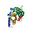
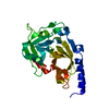
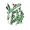
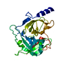
| Entry | Database: PDB / ID: 1dt2 | ||||||
|---|---|---|---|---|---|---|---|
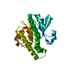
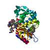
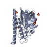
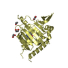
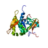
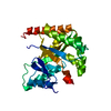
| Title | CRYSTAL STRUCTURE OF EXFOLIATIVE TOXIN B | ||||||
 Components Components | EXFOLIATIVE TOXIN B | ||||||
 Keywords Keywords | toxin / hydrolase / epidermolysis / superantigen / serine protease | ||||||
| Function / homology |  Function and homology information Function and homology informationHydrolases; Acting on peptide bonds (peptidases); Serine endopeptidases / toxin activity / serine-type endopeptidase activity / proteolysis Similarity search - Function | ||||||
| Biological species |   Staphylococcus aureus (bacteria) Staphylococcus aureus (bacteria) | ||||||
| Method |  X-RAY DIFFRACTION / SYNCHROTRON / Resolution: 2.8 Å X-RAY DIFFRACTION / SYNCHROTRON / Resolution: 2.8 Å | ||||||
 Authors Authors | Papageorgiou, A.C. / Plano, L.R.W. / Collins, C.M. / Acharya, K.R. | ||||||
 Citation Citation | Journal: Protein Sci. / Year: 2000 Title: Structural similarities and differences in Staphylococcus aureus exfoliative toxins A and B as revealed by their crystal structures. Authors: Papageorgiou, A.C. / Plano, L.R. / Collins, C.M. / Acharya, K.R. | ||||||
| History |
|
- Structure visualization
| Structure viewer | Molecule: MolmilJmol/JSmol |
|---|
- Downloads & links
-Download
| PDBx/mmCIF format | 1dt2.cif.gz | 58.5 KB | Display | PDBx/mmCIF format |
|---|---|---|---|---|
| PDB format | pdb1dt2.ent.gz | 43.4 KB | Display | PDB format |
| PDBx/mmJSON format | 1dt2.json.gz | Tree view | PDBx/mmJSON format | |
| Others |  Other downloads Other downloads |
-Validation report
| Arichive directory | https://data.pdbj.org/pub/pdb/validation_reports/dt/1dt2ftp://data.pdbj.org/pub/pdb/validation_reports/dt/1dt2 | HTTPS FTP |
|---|
-Related structure data

-Links
 PDBj
PDBj- Assembly
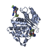
| Deposited unit | 
| ||||||||
|---|---|---|---|---|---|---|---|---|---|
| 1 |
| ||||||||
| Unit cell |
| ||||||||
| Components on special symmetry positions |
| ||||||||
| Details | The biological assembly is a monomer |
-Components
| #1: Protein | Mass: 27219.371 Da / Num. of mol.: 1 Source method: isolated from a genetically manipulated source Source: (gene. exp.) Staphylococcus aureus (bacteria)Description: CHROMOSOMAL DNA OF S. AUREUS ISOLATED FROM A PATIENT WITH STAPHYLOCOCCAL SCALDED SKIN DISEASE Plasmid: PET15B / Production host: References: UniProt: P09332, Hydrolases; Acting on peptide bonds (peptidases); Serine endopeptidases |
|---|---|
| #2: Water | ChemComp-HOH /  Mass: 18.015 Da / Num. of mol.: 9 / Source method: isolated from a natural source / Formula: H2O Mass: 18.015 Da / Num. of mol.: 9 / Source method: isolated from a natural source / Formula: H2O |
-Experimental details
-Experiment
| Experiment | Method: X-RAY DIFFRACTION / Number of used crystals: 1 |
|---|
- Sample preparation
| Crystal grow | Temperature: 289 K / Method: vapor diffusion, hanging drop / pH: 8 Details: NaFormate-no pH adjusted, so the pH should be around 8.0, VAPOR DIFFUSION, HANGING DROP, temperature 289K | |||||||||||||||
|---|---|---|---|---|---|---|---|---|---|---|---|---|---|---|---|---|
| Crystal grow | *PLUS Temperature: 16 ℃ / Method: vapor diffusion | |||||||||||||||
| Components of the solutions | *PLUS
|
-Data collection
| Diffraction | Mean temperature: 100 K |
|---|---|
| Diffraction source | Source: SYNCHROTRON / Site: EMBL/DESY, HAMBURG  / Beamline: X11 / Wavelength: 0.9057 / Beamline: X11 / Wavelength: 0.9057 |
| Detector | Type: MARRESEARCH / Detector: IMAGE PLATE / Date: Dec 4, 1998 |
| Radiation | Protocol: SINGLE WAVELENGTH / Monochromatic (M) / Laue (L): M / Scattering type: x-ray |
| Radiation wavelength | Wavelength: 0.9057 Å / Relative weight: 1 |
| Reflection | Resolution: 2.8→20 Å / Num. all: 15313 / Num. obs: 13063 / % possible obs: 85.3 % / Observed criterion σ(F): 0 / Observed criterion σ(I): -3 / Redundancy: 5.1 % / Biso Wilson estimate: 73.9 Å2 / Rmerge(I) obs: 0.083 / Net I/σ(I): 11.2 |
| Reflection shell | Resolution: 2.8→20 Å / Redundancy: 8 % / Rmerge(I) obs: 0.423 / % possible all: 69.3 |
| Reflection | *PLUS % possible obs: 85 % / Num. measured all: 66486 |
| Reflection shell | *PLUS Lowest resolution: 2.9 Å / % possible obs: 69 % / Mean I/σ(I) obs: 2.7 |
- Processing
| Software |
| ||||||||||||||||||
|---|---|---|---|---|---|---|---|---|---|---|---|---|---|---|---|---|---|---|---|
| Refinement | Resolution: 2.8→20 Å / σ(F): 0 / σ(I): -3
| ||||||||||||||||||
| Refinement step | Cycle: LAST / Resolution: 2.8→20 Å
| ||||||||||||||||||
| Refine LS restraints |
| ||||||||||||||||||
| Refinement | *PLUS % reflection Rfree: 5 % / Rfactor Rwork: 0.218 | ||||||||||||||||||
| Solvent computation | *PLUS | ||||||||||||||||||
| Displacement parameters | *PLUS | ||||||||||||||||||
| Refine LS restraints | *PLUS
|
