 Movie
Movie Controller


+ Open data

- Basic information
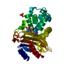
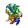
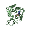
| Entry | Database: PDB / ID: 3ny4 | ||||||
|---|---|---|---|---|---|---|---|
| Title | Crystal Structure of BlaC-K73A bound with Cefamandole | ||||||
 Components Components | Beta-lactamase | ||||||
 Keywords Keywords | HYDROLASE/ANTIBIOTIC / penicillin binding protein / beta-lactam complex / HYDROLASE-ANTIBIOTIC complex | ||||||
| Function / homology |  Function and homology information Function and homology informationbeta-lactam antibiotic catabolic process / beta-lactamase activity / beta-lactamase / periplasmic space / response to antibiotic / extracellular region / plasma membrane Similarity search - Function | ||||||
| Biological species |   Mycobacterium tuberculosis (bacteria) Mycobacterium tuberculosis (bacteria) | ||||||
| Method |  X-RAY DIFFRACTION / SYNCHROTRON / MOLECULAR REPLACEMENT / Resolution: 1.22 Å X-RAY DIFFRACTION / SYNCHROTRON / MOLECULAR REPLACEMENT / Resolution: 1.22 Å | ||||||
 Authors Authors | Tremblay, L.W. / Blanchard, J.S. | ||||||
 Citation Citation | Journal: Biochemistry / Year: 2010 Title: Structures of the Michaelis Complex (1.2 A) and the Covalent Acyl Intermediate (2.0 A) of Cefamandole Bound in the Active Sites of the Mycobacterium tuberculosis beta-Lactamase K73A and E166A Mutants. Authors: Tremblay, L.W. / Xu, H. / Blanchard, J.S. | ||||||
| History |
|
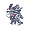
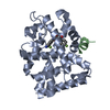
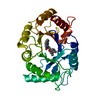
- Structure visualization
| Structure viewer | Molecule: MolmilJmol/JSmol |
|---|
- Downloads & links
-Download
| PDBx/mmCIF format | 3ny4.cif.gz | 129.5 KB | Display | PDBx/mmCIF format |
|---|---|---|---|---|
| PDB format | pdb3ny4.ent.gz | 101.2 KB | Display | PDB format |
| PDBx/mmJSON format | 3ny4.json.gz | Tree view | PDBx/mmJSON format | |
| Others |  Other downloads Other downloads |
-Validation report
| Arichive directory | https://data.pdbj.org/pub/pdb/validation_reports/ny/3ny4ftp://data.pdbj.org/pub/pdb/validation_reports/ny/3ny4 | HTTPS FTP |
|---|
-Related structure data

-Links
 PDBj
PDBj


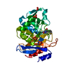
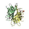
- Assembly
| Deposited unit | 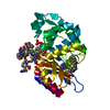
| ||||||||
|---|---|---|---|---|---|---|---|---|---|
| 1 |
| ||||||||
| Unit cell |
|
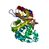
-Components
| #1: Protein | Mass: 28214.623 Da / Num. of mol.: 1 / Mutation: K87A Source method: isolated from a genetically manipulated source Source: (gene. exp.) Mycobacterium tuberculosis (bacteria) / Gene: blaA, blaC, Rv2068c, MT2128, MTCY49.07c / Plasmid: pET28a / Production host: References: UniProt: P0C5C1, UniProt: P9WKD3*PLUS, beta-lactamase | ||
|---|---|---|---|
| #2: Chemical | ChemComp-PO4 /   Mass: 94.971 Da / Num. of mol.: 1 / Source method: obtained synthetically / Formula: PO4 Mass: 94.971 Da / Num. of mol.: 1 / Source method: obtained synthetically / Formula: PO4 | ||
| #3: Chemical | ChemComp-SMX / ( 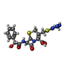  Mass: 462.503 Da / Num. of mol.: 5 / Source method: obtained synthetically / Formula: C18H18N6O5S2 / Comment: antibiotic*YM Mass: 462.503 Da / Num. of mol.: 5 / Source method: obtained synthetically / Formula: C18H18N6O5S2 / Comment: antibiotic*YM#4: Water | ChemComp-HOH / |  Mass: 18.015 Da / Num. of mol.: 237 / Source method: isolated from a natural source / Formula: H2O Mass: 18.015 Da / Num. of mol.: 237 / Source method: isolated from a natural source / Formula: H2O |
-Experimental details
-Experiment
| Experiment | Method: X-RAY DIFFRACTION / Number of used crystals: 1 |
|---|
- Sample preparation
| Crystal | Density Matthews: 2.26 Å3/Da / Density % sol: 45.66 % |
|---|---|
| Crystal grow | Temperature: 298 K / Method: vapor diffusion, sitting drop / pH: 7.5 Details: 0.1 M HEPES, 2 M NH4H2PO4, pH 7.5, Vapor diffusion, Sitting drop, temperature 298K |
-Data collection
| Diffraction | Mean temperature: 100 K |
|---|---|
| Diffraction source | Source: SYNCHROTRON / Site: NSLS  / Beamline: X29A / Wavelength: 1 Å / Beamline: X29A / Wavelength: 1 Å |
| Detector | Type: ADSC QUANTUM 210 / Detector: CCD / Date: Jun 30, 2010 |
| Radiation | Monochromator: Si(111) Channel Cut / Protocol: SINGLE WAVELENGTH / Scattering type: x-ray |
| Radiation wavelength | Wavelength: 1 Å / Relative weight: 1 |
| Reflection | Resolution: 1.22→50 Å / Num. all: 76776 / Num. obs: 76623 / % possible obs: 99.8 % / Observed criterion σ(F): 0 / Observed criterion σ(I): 0 / Redundancy: 7.6 % / Biso Wilson estimate: 10.218 Å2 / Rmerge(I) obs: 0.069 / Net I/σ(I): 32.5 |
| Reflection shell | Resolution: 1.22→1.24 Å / Redundancy: 5.9 % / Rmerge(I) obs: 0.698 / Mean I/σ(I) obs: 2.1 / Num. unique all: 3769 / % possible all: 99.1 |
- Processing
| Software |
| ||||||||||||||||||||||||||||||||||||||||||||||||||||||||||||||||||||||
|---|---|---|---|---|---|---|---|---|---|---|---|---|---|---|---|---|---|---|---|---|---|---|---|---|---|---|---|---|---|---|---|---|---|---|---|---|---|---|---|---|---|---|---|---|---|---|---|---|---|---|---|---|---|---|---|---|---|---|---|---|---|---|---|---|---|---|---|---|---|---|---|
| Refinement | Method to determine structure: MOLECULAR REPLACEMENT / Resolution: 1.22→40.16 Å / Cor.coef. Fo:Fc: 0.97 / Cor.coef. Fo:Fc free: 0.964 / Occupancy max: 1 / Occupancy min: 0.5 / SU B: 1.119 / SU ML: 0.023 / Cross valid method: THROUGHOUT / σ(F): 0 / ESU R Free: 0.04 / Stereochemistry target values: MAXIMUM LIKELIHOOD Details: HYDROGENS HAVE BEEN ADDED IN THE RIDING POSITIONS. U VALUES REFINED INDIVIDUALLY.
| ||||||||||||||||||||||||||||||||||||||||||||||||||||||||||||||||||||||
| Solvent computation | Ion probe radii: 0.8 Å / Shrinkage radii: 0.8 Å / VDW probe radii: 1.4 Å / Solvent model: MASK | ||||||||||||||||||||||||||||||||||||||||||||||||||||||||||||||||||||||
| Displacement parameters | Biso max: 104.8 Å2 / Biso mean: 14.7059 Å2 / Biso min: 5.51 Å2
| ||||||||||||||||||||||||||||||||||||||||||||||||||||||||||||||||||||||
| Refinement step | Cycle: LAST / Resolution: 1.22→40.16 Å
| ||||||||||||||||||||||||||||||||||||||||||||||||||||||||||||||||||||||
| Refine LS restraints |
| ||||||||||||||||||||||||||||||||||||||||||||||||||||||||||||||||||||||
| LS refinement shell | Resolution: 1.22→1.252 Å / Total num. of bins used: 20
|
