 Movie
Movie Controller


+ Open data

- Basic information
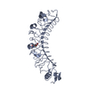
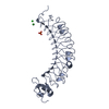
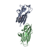
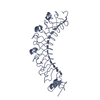
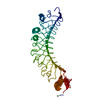
| Entry | Database: PDB / ID: 1dqv | ||||||
|---|---|---|---|---|---|---|---|
| Title | CRYSTAL STRUCTURE OF SYNAPTOTAGMIN III C2A/C2B | ||||||
 Components Components | SYNAPTOTAGMIN III | ||||||
 Keywords Keywords | ENDOCYTOSIS/EXOCYTOSIS / beta sandwich / calcium ion / C2 domain / ENDOCYTOSIS-EXOCYTOSIS COMPLEX | ||||||
| Function / homology |  Function and homology information Function and homology informationregulation of calcium ion-dependent exocytosis / calcium ion sensor activity / exocytic vesicle / positive regulation of vesicle fusion / positive regulation of dendrite extension / calcium-ion regulated exocytosis / calcium-dependent phospholipid binding / syntaxin binding / transport vesicle membrane / phosphatidylserine binding ...regulation of calcium ion-dependent exocytosis / calcium ion sensor activity / exocytic vesicle / positive regulation of vesicle fusion / positive regulation of dendrite extension / calcium-ion regulated exocytosis / calcium-dependent phospholipid binding / syntaxin binding / transport vesicle membrane / phosphatidylserine binding / clathrin binding / synaptic vesicle exocytosis / regulation of postsynaptic membrane neurotransmitter receptor levels / vesicle-mediated transport / phosphatidylinositol-4,5-bisphosphate binding / SNARE binding / synaptic membrane / response to calcium ion / presynapse / chemical synaptic transmission / cell differentiation / endosome / postsynapse / protein heterodimerization activity / calcium ion binding / protein homodimerization activity / identical protein binding / plasma membrane Similarity search - Function | ||||||
| Biological species |  | ||||||
| Method |  X-RAY DIFFRACTION / SYNCHROTRON / Resolution: 3.2 Å X-RAY DIFFRACTION / SYNCHROTRON / Resolution: 3.2 Å | ||||||
 Authors Authors | Sutton, R.B. / Ernst, J.A. / Brunger, A.T. | ||||||
 Citation Citation | Journal: J.Cell Biol. / Year: 1999 Title: Crystal structure of the cytosolic C2A-C2B domains of synaptotagmin III. Implications for Ca(+2)-independent snare complex interaction. Authors: Sutton, R.B. / Ernst, J.A. / Brunger, A.T. | ||||||
| History |
|
- Structure visualization
| Structure viewer | Molecule: MolmilJmol/JSmol |
|---|
- Downloads & links
-Download
| PDBx/mmCIF format | 1dqv.cif.gz | 66.1 KB | Display | PDBx/mmCIF format |
|---|---|---|---|---|
| PDB format | pdb1dqv.ent.gz | 49.5 KB | Display | PDB format |
| PDBx/mmJSON format | 1dqv.json.gz | Tree view | PDBx/mmJSON format | |
| Others |  Other downloads Other downloads |
-Validation report
| Arichive directory | https://data.pdbj.org/pub/pdb/validation_reports/dq/1dqvftp://data.pdbj.org/pub/pdb/validation_reports/dq/1dqv | HTTPS FTP |
|---|
-Related structure data
| Related structure data | |
|---|---|
| Similar structure data |


-Links
 PDBj
PDBj


- Assembly
| Deposited unit | 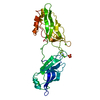
| ||||||||
|---|---|---|---|---|---|---|---|---|---|
| 1 |
| ||||||||
| 2 | 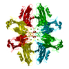
| ||||||||
| Unit cell |
| ||||||||
| Details | The biological assembly is one C2A domain and one C2B domain |
-Components
| #1: Protein | Mass: 33253.914 Da / Num. of mol.: 1 / Fragment: C2A/C2B Source method: isolated from a genetically manipulated source Details: SOLUBLE PORTION / Source: (gene. exp.)  | ||
|---|---|---|---|
| #2: Chemical | ChemComp-MG /   Mass: 24.305 Da / Num. of mol.: 4 / Source method: obtained synthetically / Formula: Mg Mass: 24.305 Da / Num. of mol.: 4 / Source method: obtained synthetically / Formula: Mg#3: Chemical | ChemComp-SO4 / |   Mass: 96.063 Da / Num. of mol.: 1 / Source method: obtained synthetically / Formula: SO4 Mass: 96.063 Da / Num. of mol.: 1 / Source method: obtained synthetically / Formula: SO4 |
-Experimental details
-Experiment
| Experiment | Method: X-RAY DIFFRACTION / Number of used crystals: 11 |
|---|
- Sample preparation
| Crystal | Density Matthews: 4.08 Å3/Da / Density % sol: 69.82 % | ||||||||||||||||||||||||
|---|---|---|---|---|---|---|---|---|---|---|---|---|---|---|---|---|---|---|---|---|---|---|---|---|---|
| Crystal grow | Temperature: 293 K / Method: vapor diffusion, hanging drop / pH: 6.5 Details: 1.6M Magnesium Sulfate, pH 6.5, VAPOR DIFFUSION, HANGING DROP, temperature 293K | ||||||||||||||||||||||||
| Crystal grow | *PLUS Temperature: 20 ℃ | ||||||||||||||||||||||||
| Components of the solutions | *PLUS
|
-Data collection
| Diffraction | Mean temperature: 293 K |
|---|---|
| Diffraction source | Source: SYNCHROTRON / Site: SSRL  / Beamline: BL1-5 / Wavelength: 1 / Beamline: BL1-5 / Wavelength: 1 |
| Detector | Type: ADSC QUANTUM 4 / Detector: CCD / Date: Mar 13, 1998 |
| Radiation | Protocol: SINGLE WAVELENGTH / Monochromatic (M) / Laue (L): M / Scattering type: x-ray |
| Radiation wavelength | Wavelength: 1 Å / Relative weight: 1 |
| Reflection | Resolution: 3.2→50 Å / Num. all: 61080 / Num. obs: 9578 / % possible obs: 99.6 % / Observed criterion σ(F): 3 / Observed criterion σ(I): 3 / Redundancy: 6.3 % / Biso Wilson estimate: -1 Å2 / Rmerge(I) obs: 0.05 / Net I/σ(I): 2.9 |
| Reflection shell | Resolution: 3.2→3.31 Å / Redundancy: 7 % / Rmerge(I) obs: 0.785 / Num. unique all: 929 / % possible all: 99.9 |
| Reflection | *PLUS Num. measured all: 61080 |
| Reflection shell | *PLUS % possible obs: 99.9 % |
- Processing
| Software |
| |||||||||||||||||||||||||
|---|---|---|---|---|---|---|---|---|---|---|---|---|---|---|---|---|---|---|---|---|---|---|---|---|---|---|
| Refinement | Resolution: 3.2→50 Å / σ(F): 0 / σ(I): 0 / Stereochemistry target values: Engh & Huber Details: used maximum likelihood/experiment phase set target function
| |||||||||||||||||||||||||
| Refinement step | Cycle: LAST / Resolution: 3.2→50 Å
| |||||||||||||||||||||||||
| Refine LS restraints |
| |||||||||||||||||||||||||
| Software | *PLUS Name: 'CNS' / Classification: refinement | |||||||||||||||||||||||||
| Refinement | *PLUS Rfactor Rfree: 0.349 | |||||||||||||||||||||||||
| Solvent computation | *PLUS | |||||||||||||||||||||||||
| Displacement parameters | *PLUS | |||||||||||||||||||||||||
| Refine LS restraints | *PLUS
| |||||||||||||||||||||||||
| LS refinement shell | *PLUS Highest resolution: 3.2 Å / Lowest resolution: 3.45 Å / Rfactor Rfree: 0.344 / Rfactor Rwork: 0.302 |
