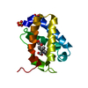
 Movie
Movie Controller


+ Open data

- Basic information
| Entry | Database: PDB / ID: 1dlw | ||||||
|---|---|---|---|---|---|---|---|
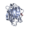
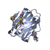
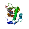
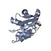
| Title | X-RAY CRYSTAL STRUCTURE OF TRUNCATED HEMOGLOBIN FROM P.CAUDATUM. | ||||||
 Components Components | HEMOGLOBIN | ||||||
 Keywords Keywords | OXYGEN STORAGE/TRANSPORT / GLOBIN FOLD TRUNCATED HEMOGLOBIN NON VERTEBRATE HEMOGLOBIN / OXYGEN STORAGE-TRANSPORT COMPLEX | ||||||
| Function / homology |  Function and homology information Function and homology informationoxygen carrier activity / oxygen binding / heme binding / metal ion binding Similarity search - Function | ||||||
| Biological species |  | ||||||
| Method |  X-RAY DIFFRACTION / SYNCHROTRON / Resolution: 1.54 Å X-RAY DIFFRACTION / SYNCHROTRON / Resolution: 1.54 Å | ||||||
 Authors Authors | Pesce, A. / Couture, M. / Guertin, M. / Dewilde, S. / Moens, L. / Bolognesi, M. | ||||||
 Citation Citation | Journal: EMBO J. / Year: 2000 Title: A novel two-over-two alpha-helical sandwich fold is characteristic of the truncated hemoglobin family. Authors: Pesce, A. / Couture, M. / Dewilde, S. / Guertin, M. / Yamauchi, K. / Ascenzi, P. / Moens, L. / Bolognesi, M. | ||||||
| History |
|
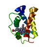
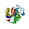
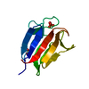
- Structure visualization
| Structure viewer | Molecule: MolmilJmol/JSmol |
|---|
- Downloads & links
-Download
| PDBx/mmCIF format | 1dlw.cif.gz | 65.9 KB | Display | PDBx/mmCIF format |
|---|---|---|---|---|
| PDB format | pdb1dlw.ent.gz | 47.7 KB | Display | PDB format |
| PDBx/mmJSON format | 1dlw.json.gz | Tree view | PDBx/mmJSON format | |
| Others |  Other downloads Other downloads |
-Validation report
| Arichive directory | https://data.pdbj.org/pub/pdb/validation_reports/dl/1dlwftp://data.pdbj.org/pub/pdb/validation_reports/dl/1dlw | HTTPS FTP |
|---|
-Related structure data


-Links
 PDBj
PDBj


- Assembly
| Deposited unit | 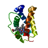
| ||||||||
|---|---|---|---|---|---|---|---|---|---|
| 1 |
| ||||||||
| Unit cell |
|
-Components
| #1: Protein | Mass: 11914.342 Da / Num. of mol.: 1 Source method: isolated from a genetically manipulated source Source: (gene. exp.)  |
|---|---|
| #2: Chemical | ChemComp-HEM /   Mass: 616.487 Da / Num. of mol.: 1 / Source method: obtained synthetically / Formula: C34H32FeN4O4 Mass: 616.487 Da / Num. of mol.: 1 / Source method: obtained synthetically / Formula: C34H32FeN4O4 |
| #3: Water | ChemComp-HOH /  Mass: 18.015 Da / Num. of mol.: 207 / Source method: isolated from a natural source / Formula: H2O Mass: 18.015 Da / Num. of mol.: 207 / Source method: isolated from a natural source / Formula: H2O |
| Compound details | TRUNCATED HEMOGLOBIN |
-Experimental details
-Experiment
| Experiment | Method: X-RAY DIFFRACTION / Number of used crystals: 1 |
|---|
- Sample preparation
| Crystal | Density Matthews: 2.81 Å3/Da / Density % sol: 56.23 % | |||||||||||||||||||||||||
|---|---|---|---|---|---|---|---|---|---|---|---|---|---|---|---|---|---|---|---|---|---|---|---|---|---|---|
| Crystal grow | Temperature: 298 K / Method: vapor diffusion, hanging drop / pH: 6 Details: AMMONIUM SULPHATE, SODIUM ACETATE, pH 6.0, VAPOR DIFFUSION, HANGING DROP, temperature 298K | |||||||||||||||||||||||||
| Crystal grow | *PLUS Temperature: 4 ℃ / Method: vapor diffusion / pH: 5.5 | |||||||||||||||||||||||||
| Components of the solutions | *PLUS
|
-Data collection
| Diffraction | Mean temperature: 100 K |
|---|---|
| Diffraction source | Source: SYNCHROTRON / Site: ESRF  / Beamline: BM14 / Wavelength: 1 / Beamline: BM14 / Wavelength: 1 |
| Detector | Type: MARRESEARCH / Detector: IMAGE PLATE / Date: Feb 19, 1999 |
| Radiation | Protocol: SINGLE WAVELENGTH / Monochromatic (M) / Laue (L): M / Scattering type: x-ray |
| Radiation wavelength | Wavelength: 1 Å / Relative weight: 1 |
| Reflection | Resolution: 1.54→30 Å / Num. all: 36762 / Num. obs: 36762 / % possible obs: 96.7 % / Observed criterion σ(F): 0 / Observed criterion σ(I): 0 / Redundancy: 3 % / Biso Wilson estimate: 12 Å2 / Rmerge(I) obs: 0.025 / Net I/σ(I): 31 |
| Reflection shell | Resolution: 1.54→1.57 Å / Redundancy: 2 % / Rmerge(I) obs: 0.106 / % possible all: 93.9 |
| Reflection | *PLUS |
| Reflection shell | *PLUS % possible obs: 88.2 % / Mean I/σ(I) obs: 7 |
- Processing
| Software |
| ||||||||||||||||||||||||||||||||||||||||||||||||||||||||||||
|---|---|---|---|---|---|---|---|---|---|---|---|---|---|---|---|---|---|---|---|---|---|---|---|---|---|---|---|---|---|---|---|---|---|---|---|---|---|---|---|---|---|---|---|---|---|---|---|---|---|---|---|---|---|---|---|---|---|---|---|---|---|
| Refinement | Resolution: 1.54→30 Å / σ(F): 0 / σ(I): 0 / Stereochemistry target values: ENGH & HUBER Details: AT THE END OF REFINEMENT, THE ANISOTROPIC REFINEMENT OF B-FACTORS WAS USED.
| ||||||||||||||||||||||||||||||||||||||||||||||||||||||||||||
| Refinement step | Cycle: LAST / Resolution: 1.54→30 Å
| ||||||||||||||||||||||||||||||||||||||||||||||||||||||||||||
| Refine LS restraints |
| ||||||||||||||||||||||||||||||||||||||||||||||||||||||||||||
| Software | *PLUS Name: CNS / Classification: refinement | ||||||||||||||||||||||||||||||||||||||||||||||||||||||||||||
| Refinement | *PLUS Lowest resolution: 30 Å / σ(F): 0 | ||||||||||||||||||||||||||||||||||||||||||||||||||||||||||||
| Solvent computation | *PLUS | ||||||||||||||||||||||||||||||||||||||||||||||||||||||||||||
| Displacement parameters | *PLUS |
