 Movie
Movie Controller

[English] 日本語
 Yorodumi
- PDB-1dl7: THE STRUCTURAL BASIS OF REPERTOIRE SHIFT IN AN IMMUNE RESPONSE TO... -
+ Open data

- Basic information
| Entry | Database: PDB / ID: 1dl7 | ||||||
|---|---|---|---|---|---|---|---|
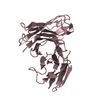
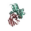
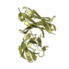
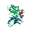
| Title | THE STRUCTURAL BASIS OF REPERTOIRE SHIFT IN AN IMMUNE RESPONSE TO PHOSPHOCHOLINE | ||||||
 Components Components |
| ||||||
 Keywords Keywords | IMMUNE SYSTEM / SINGLE CHAIN FV / REPERTOIRE SHIFT | ||||||
| Function / homology |  Function and homology information Function and homology informationimmunoglobulin mediated immune response / immunoglobulin complex / antigen binding / adaptive immune response / immune response / extracellular region Similarity search - Function | ||||||
| Biological species |  | ||||||
| Method |  X-RAY DIFFRACTION / Resolution: 2.35 Å X-RAY DIFFRACTION / Resolution: 2.35 Å | ||||||
 Authors Authors | Schumacher, M. / Brown, M. | ||||||
 Citation Citation | Journal: J.Exp.Med. / Year: 2000 Title: The structural basis of repertoire shift in an immune response to phosphocholine. Authors: Brown, M. / Schumacher, M.A. / Wiens, G.D. / Brennan, R.G. / Rittenberg, M.B. | ||||||
| History |
|
- Structure visualization
| Structure viewer | Molecule: MolmilJmol/JSmol |
|---|
- Downloads & links
-Download
| PDBx/mmCIF format | 1dl7.cif.gz | 57.4 KB | Display | PDBx/mmCIF format |
|---|---|---|---|---|
| PDB format | pdb1dl7.ent.gz | 41.1 KB | Display | PDB format |
| PDBx/mmJSON format | 1dl7.json.gz | Tree view | PDBx/mmJSON format | |
| Others |  Other downloads Other downloads |
-Validation report
| Arichive directory | https://data.pdbj.org/pub/pdb/validation_reports/dl/1dl7ftp://data.pdbj.org/pub/pdb/validation_reports/dl/1dl7 | HTTPS FTP |
|---|
-Related structure data
| Similar structure data |
|---|


-Links
 PDBj
PDBj

- Assembly
| Deposited unit | 
| ||||||||
|---|---|---|---|---|---|---|---|---|---|
| 1 |
| ||||||||
| Unit cell |
| ||||||||
| Atom site foot note | 1: CIS PROLINE - PRO H 101 | ||||||||
| Details | the biological assembly is a dimer of one light chain and one heavy chain. |
-Components
| #1: Antibody | Mass: 11544.834 Da / Num. of mol.: 1 / Fragment: FV (SINGLE CHAIN) Source method: isolated from a genetically manipulated source Details: COMBINED LIGHT AND HEAVY CHAIN VIA (G4S)3 LINKER / Source: (gene. exp.)  |
|---|---|
| #2: Antibody | Mass: 12333.841 Da / Num. of mol.: 1 / Fragment: FV (SINGLE CHAIN) Source method: isolated from a genetically manipulated source Source: (gene. exp.) |
| #3: Chemical | ChemComp-NCH /   Mass: 305.244 Da / Num. of mol.: 1 / Source method: obtained synthetically / Formula: C11H18N2O6P Mass: 305.244 Da / Num. of mol.: 1 / Source method: obtained synthetically / Formula: C11H18N2O6P |
| #4: Water | ChemComp-HOH /  Mass: 18.015 Da / Num. of mol.: 79 / Source method: isolated from a natural source / Formula: H2O Mass: 18.015 Da / Num. of mol.: 79 / Source method: isolated from a natural source / Formula: H2O |
| Has protein modification | Y |
-Experimental details
-Experiment
| Experiment | Method: X-RAY DIFFRACTION / Number of used crystals: 1 |
|---|
- Sample preparation
| Crystal | Density Matthews: 2.48 Å3/Da / Density % sol: 50.43 % | ||||||||||||||||||||||||||||||
|---|---|---|---|---|---|---|---|---|---|---|---|---|---|---|---|---|---|---|---|---|---|---|---|---|---|---|---|---|---|---|---|
| Crystal grow | Temperature: 298 K / Method: vapor diffusion, hanging drop / pH: 6.5 Details: SODIUM/POTASSIUM PHOSPHATE, HEPES, pH 6.5, VAPOR DIFFUSION, HANGING DROP, temperature 298.K | ||||||||||||||||||||||||||||||
| Crystal grow | *PLUS Method: vapor diffusion | ||||||||||||||||||||||||||||||
| Components of the solutions | *PLUS
|
-Data collection
| Diffraction | Mean temperature: 298 K |
|---|---|
| Diffraction source | Source: ROTATING ANODE / Type: RIGAKU RU300 / Wavelength: 1.5418 |
| Radiation | Protocol: SINGLE WAVELENGTH / Monochromatic (M) / Laue (L): M / Scattering type: x-ray |
| Radiation wavelength | Wavelength: 1.5418 Å / Relative weight: 1 |
| Reflection | Resolution: 2.35→10 Å / Num. all: 47930 / Num. obs: 8983 / % possible obs: 84 % / Observed criterion σ(F): 1 / Observed criterion σ(I): 1 / Redundancy: 2 % / Biso Wilson estimate: 20 Å2 / Rmerge(I) obs: 0.073 / Net I/σ(I): 8.5 |
| Reflection shell | Resolution: 2.35→2.42 Å / Redundancy: 2 % / Rmerge(I) obs: 0.3 / % possible all: 52 |
| Reflection | *PLUS Num. measured all: 47930 |
| Reflection shell | *PLUS % possible obs: 47 % / Rmerge(I) obs: 0.265 / Mean I/σ(I) obs: 1.5 |
- Processing
| Software |
| ||||||||||||||||||||||||||||||
|---|---|---|---|---|---|---|---|---|---|---|---|---|---|---|---|---|---|---|---|---|---|---|---|---|---|---|---|---|---|---|---|
| Refinement | Resolution: 2.35→10 Å / σ(F): 1 / σ(I): 1 / Stereochemistry target values: ENGH AND HUBER
| ||||||||||||||||||||||||||||||
| Refinement step | Cycle: LAST / Resolution: 2.35→10 Å
| ||||||||||||||||||||||||||||||
| Refine LS restraints |
| ||||||||||||||||||||||||||||||
| Software | *PLUS Name: TNT / Classification: refinement | ||||||||||||||||||||||||||||||
| Refinement | *PLUS Rfactor Rwork: 0.191 | ||||||||||||||||||||||||||||||
| Solvent computation | *PLUS | ||||||||||||||||||||||||||||||
| Displacement parameters | *PLUS |
