 Movie
Movie Controller

[English] 日本語
 Yorodumi
- PDB-1df5: INTERACTIONS BETWEEN HIV-1 GP41 CORE AND DETERGENTS AND THEIR IMP... -
+ Open data

- Basic information
| Entry | Database: PDB / ID: 1df5 | ||||||
|---|---|---|---|---|---|---|---|
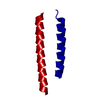
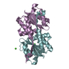
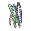
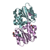
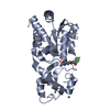
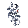
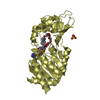
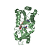
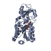
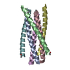
| Title | INTERACTIONS BETWEEN HIV-1 GP41 CORE AND DETERGENTS AND THEIR IMPLICATIONS FOR MEMBRANE FUSION | ||||||
 Components Components | HIV-1 ENVELOPE GLYCOPROTEIN GP41 | ||||||
 Keywords Keywords | VIRAL PROTEIN / HIV-1 / GP41 / MEMBRANE FUSION / PROTEIN-DETERGENT INTERACTION | ||||||
| Function / homology |  Function and homology information Function and homology informationSynthesis and processing of ENV and VPU / symbiont-mediated evasion of host immune response / positive regulation of establishment of T cell polarity / Alpha-defensins / Dectin-2 family / Binding and entry of HIV virion / positive regulation of plasma membrane raft polarization / positive regulation of receptor clustering / host cell endosome membrane / actin filament organization ...Synthesis and processing of ENV and VPU / symbiont-mediated evasion of host immune response / positive regulation of establishment of T cell polarity / Alpha-defensins / Dectin-2 family / Binding and entry of HIV virion / positive regulation of plasma membrane raft polarization / positive regulation of receptor clustering / host cell endosome membrane / actin filament organization / Assembly Of The HIV Virion / Budding and maturation of HIV virion / clathrin-dependent endocytosis of virus by host cell / viral protein processing / receptor ligand activity / fusion of virus membrane with host plasma membrane / fusion of virus membrane with host endosome membrane / viral envelope / symbiont entry into host cell / virion attachment to host cell / host cell plasma membrane / virion membrane / structural molecule activity / membrane Similarity search - Function | ||||||
| Biological species |   Human immunodeficiency virus 1 Human immunodeficiency virus 1 | ||||||
| Method |  X-RAY DIFFRACTION / Resolution: 2.7 Å X-RAY DIFFRACTION / Resolution: 2.7 Å | ||||||
 Authors Authors | Shu, W. / Ji, H. / Lu, M. | ||||||
 Citation Citation | Journal: J.Biol.Chem. / Year: 2000 Title: Interactions between HIV-1 gp41 core and detergents and their implications for membrane fusion. Authors: Shu, W. / Ji, H. / Lu, M. | ||||||
| History |
|
- Structure visualization
| Structure viewer | Molecule: MolmilJmol/JSmol |
|---|
- Downloads & links
-Download
| PDBx/mmCIF format | 1df5.cif.gz | 23.9 KB | Display | PDBx/mmCIF format |
|---|---|---|---|---|
| PDB format | pdb1df5.ent.gz | 16.1 KB | Display | PDB format |
| PDBx/mmJSON format | 1df5.json.gz | Tree view | PDBx/mmJSON format | |
| Others |  Other downloads Other downloads |
-Validation report
| Arichive directory | https://data.pdbj.org/pub/pdb/validation_reports/df/1df5ftp://data.pdbj.org/pub/pdb/validation_reports/df/1df5 | HTTPS FTP |
|---|
-Related structure data

-Links
 PDBj
PDBj


- Assembly
| Deposited unit | 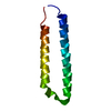
| ||||||||
|---|---|---|---|---|---|---|---|---|---|
| 1 | 
| ||||||||
| Unit cell |
| ||||||||
| Details | The biological assembly is a homotrimer. |
-Components
| #1: Protein | Mass: 7879.753 Da / Num. of mol.: 1 Fragment: RESIDUES 1 - 34 AND 41 - 68 CONNECTED BY A SIX-RESIDUE LINKER (SER-GLY-GLY-ARG-GLY-GLY) Source method: isolated from a genetically manipulated source Source: (gene. exp.) Human immunodeficiency virus 1 / Genus: LentivirusDescription: RECOMBINANT GP41 WITH LINKER (SER-GLY-GLY- ARG-GLY-GLY) BETWEEN TWO FRAGMENTS Production host:  |
|---|---|
| Compound details | IN THE STRUCTURE, SEQUENCE 1 - 34 IS FROM GP41 RESIDUES 546 - 579 (IN GP160 NUMBERING SYSTEM), 35 - ...IN THE STRUCTURE, SEQUENCE 1 - 34 IS FROM GP41 RESIDUES 546 - 579 (IN GP160 NUMBERING SYSTEM), 35 - 40 IS AN ARTIFICIAL |
-Experimental details
-Experiment
| Experiment | Method: X-RAY DIFFRACTION / Number of used crystals: 1 |
|---|
- Sample preparation
| Crystal | Density Matthews: 2.04 Å3/Da / Density % sol: 39.64 % | |||||||||||||||||||||||||
|---|---|---|---|---|---|---|---|---|---|---|---|---|---|---|---|---|---|---|---|---|---|---|---|---|---|---|
| Crystal grow | Temperature: 293 K / Method: vapor diffusion, hanging drop / pH: 7.5 Details: Potassium sodium tartrate, SDS, sodium hepes, pH 7.5, VAPOR DIFFUSION, HANGING DROP, temperature 293K | |||||||||||||||||||||||||
| Crystal grow | *PLUS | |||||||||||||||||||||||||
| Components of the solutions | *PLUS
|
-Data collection
| Diffraction | Mean temperature: 130 K |
|---|---|
| Diffraction source | Source: ROTATING ANODE / Type: RIGAKU RU200 / Wavelength: 1.5418 |
| Detector | Type: RIGAKU RAXIS IV / Detector: IMAGE PLATE / Date: Mar 19, 1998 |
| Radiation | Protocol: SINGLE WAVELENGTH / Monochromatic (M) / Laue (L): M / Scattering type: x-ray |
| Radiation wavelength | Wavelength: 1.5418 Å / Relative weight: 1 |
| Reflection | Resolution: 2.7→30 Å / Num. all: 1859 / Num. obs: 1859 / % possible obs: 99.9 % / Observed criterion σ(F): 0 / Observed criterion σ(I): 0 / Redundancy: 5.6 % / Rmerge(I) obs: 0.046 / Net I/σ(I): 21 |
| Reflection shell | Resolution: 2.7→2.8 Å / Redundancy: 5.8 % / Rmerge(I) obs: 0.102 / Num. unique all: 186 / % possible all: 100 |
| Reflection | *PLUS Num. measured all: 10354 |
| Reflection shell | *PLUS % possible obs: 100 % |
- Processing
| Software |
| |||||||||||||||||||||||||
|---|---|---|---|---|---|---|---|---|---|---|---|---|---|---|---|---|---|---|---|---|---|---|---|---|---|---|
| Refinement | Resolution: 2.7→8 Å / σ(F): 0 / σ(I): 0 / Stereochemistry target values: Engh & Huber
| |||||||||||||||||||||||||
| Refinement step | Cycle: LAST / Resolution: 2.7→8 Å
| |||||||||||||||||||||||||
| Refine LS restraints |
| |||||||||||||||||||||||||
| Software | *PLUS Name: X-PLOR / Version: 3.1 / Classification: refinement | |||||||||||||||||||||||||
| Refine LS restraints | *PLUS
|
