 ムービー
ムービー コントローラー


+ データを開く

- 基本情報
基本情報
| 登録情報 | データベース: PDB / ID: 1deq | ||||||
|---|---|---|---|---|---|---|---|
| タイトル | THE CRYSTAL STRUCTURE OF MODIFIED BOVINE FIBRINOGEN (AT ~4 ANGSTROM RESOLUTION) | ||||||
 要素 要素 |
| ||||||
 キーワード キーワード | BLOOD CLOTTING / COILED-COIL | ||||||
| 機能・相同性 |  機能・相同性情報 機能・相同性情報blood coagulation, common pathway / fibrinogen complex / blood coagulation, fibrin clot formation / positive regulation of heterotypic cell-cell adhesion / protein polymerization / fibrinolysis / cell-matrix adhesion / platelet aggregation / : / protein-macromolecule adaptor activity ...blood coagulation, common pathway / fibrinogen complex / blood coagulation, fibrin clot formation / positive regulation of heterotypic cell-cell adhesion / protein polymerization / fibrinolysis / cell-matrix adhesion / platelet aggregation / : / protein-macromolecule adaptor activity / adaptive immune response / signaling receptor binding / innate immune response / extracellular space / metal ion binding 類似検索 - 分子機能 | ||||||
| 生物種 |  | ||||||
| 手法 |  X線回折 / シンクロトロン / molecular replacement (using human fragment d coordinates) / 解像度: 3.5 Å X線回折 / シンクロトロン / molecular replacement (using human fragment d coordinates) / 解像度: 3.5 Å | ||||||
 データ登録者 データ登録者 | Brown, J.H. / Volkmann, N. / Jun, G. / Henschen-Edman, A.H. / Cohen, C. | ||||||
 引用 引用 | ジャーナル: Proc.Natl.Acad.Sci.USA / 年: 2000 タイトル: The crystal structure of modified bovine fibrinogen. 著者: Brown, J.H. / Volkmann, N. / Jun, G. / Henschen-Edman, A.H. / Cohen, C. #1: ジャーナル: J.Mol.Biol. / 年: 1991タイトル: Fibrinogen Structure in Projection at 18 Angstroms Resolution 著者: Rao, S.P.S. / Poojary, M.D. / Elliott Jr., B.W. / Melanson, L.A. / Oriel, B. / Cohen, C. #2: ジャーナル: J.Mol.Biol. / 年: 1978タイトル: Crystals of Modified Fibrinogen: Size, Shape and Packing of Molecules 著者: Weisel, J.W. / Warren, S.G. / Cohen, C. #3: ジャーナル: J.Mol.Biol. / 年: 1977タイトル: Crystalline States of a Modified Fibrinogen 著者: Tooney, N.M. / Cohen, C. | ||||||
| 履歴 |
|
- 構造の表示
構造の表示
| 構造ビューア | 分子: MolmilJmol/JSmol |
|---|
- ダウンロードとリンク
-ダウンロード
| PDBx/mmCIF形式 | 1deq.cif.gz | 161.2 KB | 表示 | PDBx/mmCIF形式 |
|---|---|---|---|---|
| PDB形式 | pdb1deq.ent.gz | 89.3 KB | 表示 | PDB形式 |
| PDBx/mmJSON形式 | 1deq.json.gz | ツリー表示 | PDBx/mmJSON形式 | |
| その他 |  その他のダウンロード その他のダウンロード |
-検証レポート
| 文書・要旨 | 1deq_validation.pdf.gz | 406.6 KB | 表示 | wwPDB検証レポート |
|---|---|---|---|---|
| 文書・詳細版 | 1deq_full_validation.pdf.gz | 408.3 KB | 表示 | |
| XML形式データ | 1deq_validation.xml.gz | 2.4 KB | 表示 | |
| CIF形式データ | 1deq_validation.cif.gz | 37.3 KB | 表示 | |
| アーカイブディレクトリ | https://data.pdbj.org/pub/pdb/validation_reports/de/1deqftp://data.pdbj.org/pub/pdb/validation_reports/de/1deq | HTTPS FTP |
-関連構造データ


-リンク
 PDBj
PDBj


- 集合体
集合体
| 登録構造単位 | 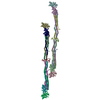
| ||||||||
|---|---|---|---|---|---|---|---|---|---|
| 1 | 
| ||||||||
| 2 | 
| ||||||||
| 単位格子 |
| ||||||||
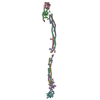
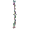
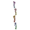
| 詳細 | An ~ 4 Angstrom structure of the 285 kDa major fragment of bovine fibrinogen. The alpha-carbon coordinates for both molecules per asymmetric unit are provided. The molecule is a dimer of a heterotrimer. Coordinates with chain ids ABC:DEF and NOP:QRS are the two crystallographically independent dimers. A,D,N, and Q correspond to the A-alpha chain. B,E,O, and R correspond to the B-beta chain. C,F,P, and S correspond to the gamma chain. These alpha-carbon coordinates include the end gamma and beta domains (i.e. the C-terminal globular domains of the gamma and B-beta chains, respectively) and the coiled coils (which are made of the A-alpha, B-beta, and gamma chains.). The density corresponding to the central disulfide knot region is too irregular to be traced at this resolution, and only some coordinates for part of this domain are included where density is seen (designated M and Z for the for the two molecules per a.u.). |
-要素
| #1: タンパク質 | 分子量: 42767.410 Da / 分子数: 4 / 断片: PSEUDOMONAS AERUGINOSA PS-1-MODIFIED FRAGMENT / 由来タイプ: 天然 / 由来: (天然) #2: タンパク質 | 分子量: 46901.641 Da / 分子数: 4 / 断片: PSEUDOMONAS AERUGINOSA PS-1-MODIFIED FRAGMENT / 由来タイプ: 天然 / 由来: (天然) #3: タンパク質 | 分子量: 46626.605 Da / 分子数: 4 / 断片: PSEUDOMONAS AERUGINOSA PS-1-MODIFIED FRAGMENT / 由来タイプ: 天然 / 由来: (天然) #4: タンパク質 | 分子量: 7677.455 Da / 分子数: 2 / 断片: PSEUDOMONAS AERUGINOSA PS-1-MODIFIED FRAGMENT / 由来タイプ: 天然 / 詳細: DISORDERED DISULFIDE KNOT REGION / 由来: (天然) |
|---|
-実験情報
-実験
| 実験 | 手法: X線回折 / 使用した結晶の数: 3 |
|---|
- 試料調製
試料調製
| 結晶 | マシュー密度: 3.12 Å3/Da / 溶媒含有率: 60.53 % | ||||||||||||||||||||||||||||||
|---|---|---|---|---|---|---|---|---|---|---|---|---|---|---|---|---|---|---|---|---|---|---|---|---|---|---|---|---|---|---|---|
| 結晶化 | 温度: 277 K / pH: 6.2 詳細: 10 mM MES, 5mM sodium azide, 2mM calcium chloride , pH 6.2, temperature 277K | ||||||||||||||||||||||||||||||
| 結晶化 | *PLUS 手法: batch method | ||||||||||||||||||||||||||||||
| 溶液の組成 | *PLUS
|
-データ収集
| 回折 |
| ||||||||||||||||||||
|---|---|---|---|---|---|---|---|---|---|---|---|---|---|---|---|---|---|---|---|---|---|
| 放射光源 |
| ||||||||||||||||||||
| 検出器 |
| ||||||||||||||||||||
| 放射 | プロトコル: SINGLE WAVELENGTH / 単色(M)・ラウエ(L): M / 散乱光タイプ: x-ray | ||||||||||||||||||||
| 放射波長 |
| ||||||||||||||||||||
| 反射 | 解像度: 3.4→210 Å / Num. all: 79465 / Num. obs: 79465 / % possible obs: 78.5 % / Observed criterion σ(F): 0 / Observed criterion σ(I): 0 / 冗長度: 4.42 % / Rmerge(I) obs: 0.075 / Net I/σ(I): 7.05 | ||||||||||||||||||||
| 反射 シェル | 解像度: 3.38→3.57 Å / 冗長度: 2.3 % / Rmerge(I) obs: 0.28 / Num. unique all: 6830 / % possible all: 46.7 | ||||||||||||||||||||
| 反射 | *PLUS | ||||||||||||||||||||
| 反射 シェル | *PLUS % possible obs: 46.7 % |

- 解析
解析
| ソフトウェア |
| |||||||||||||||||||||||||
|---|---|---|---|---|---|---|---|---|---|---|---|---|---|---|---|---|---|---|---|---|---|---|---|---|---|---|
| 精密化 | 構造決定の手法: molecular replacement (using human fragment d coordinates) 開始モデル: human fragment d coordinates (see related entry 1fza) 解像度: 3.5→10 Å / σ(F): 0 立体化学のターゲット値: modified CHARMM in X-Plor 詳細: Side chains were included in refinement. At this resolution, however, the correct side chain conformations cannot be generally determined, and only alpha Carbons are deposited here. A B- ...詳細: Side chains were included in refinement. At this resolution, however, the correct side chain conformations cannot be generally determined, and only alpha Carbons are deposited here. A B-factor equal to 999 identifies a residue whose density is generally relatively disordered and thus is not included in refinement (but generally positioned using information from non- crystallographically-related copies). Note that of the asymmetric unit's four half-dimers, that designated by chain ids N, O, and P has the most residues included in the refinement, and its electron density is best ordered. The central disulphide knot region could not be traced, and only some coordinates for part of this domain are included (resname=UNK, chainid M and Z for the two molecules per a.u.). Due to anisotropy in the the diffraction, the data in the highest shells (esp. 4.0-3.5) are quite incomplete, and hence the structure is judged overall to at ~4.0 angstroms resolution.
| |||||||||||||||||||||||||
| 精密化ステップ | サイクル: LAST / 解像度: 3.5→10 Å
| |||||||||||||||||||||||||
| 拘束条件 |
| |||||||||||||||||||||||||
| ソフトウェア | *PLUS 名称: X-PLOR / バージョン: 3.851 / 分類: refinement | |||||||||||||||||||||||||
| 拘束条件 | *PLUS
|
