[English] 日本語
 Yorodumi
- PDB-1m1j: Crystal structure of native chicken fibrinogen with two different... -
+ Open data
- Basic information
| Entry | Database: PDB / ID: 1m1j | ||||||||||||
|---|---|---|---|---|---|---|---|---|---|---|---|---|---|
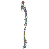
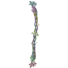
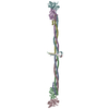
| Title | Crystal structure of native chicken fibrinogen with two different bound ligands | ||||||||||||
 Components Components |
| ||||||||||||
 Keywords Keywords | BLOOD CLOTTING / COILED COILS / DISULFIDE RINGS / FIBRINOGEN | ||||||||||||
| Function / homology |  Function and homology information Function and homology informationblood coagulation, common pathway / fibrinogen complex / blood coagulation, fibrin clot formation / positive regulation of heterotypic cell-cell adhesion / protein polymerization / fibrinolysis / cell-matrix adhesion / platelet activation / platelet aggregation / : ...blood coagulation, common pathway / fibrinogen complex / blood coagulation, fibrin clot formation / positive regulation of heterotypic cell-cell adhesion / protein polymerization / fibrinolysis / cell-matrix adhesion / platelet activation / platelet aggregation / : / protein-macromolecule adaptor activity / signaling receptor binding / extracellular space / metal ion binding Similarity search - Function | ||||||||||||
| Biological species |  | ||||||||||||
| Method |  X-RAY DIFFRACTION / X-RAY DIFFRACTION /  SYNCHROTRON / SYNCHROTRON /  MOLECULAR REPLACEMENT / Resolution: 2.7 Å MOLECULAR REPLACEMENT / Resolution: 2.7 Å | ||||||||||||
 Authors Authors | Yang, Z. / Kollman, J.M. / Pandi, L. / Doolittle, R.F. | ||||||||||||
 Citation Citation |  Journal: Biochemistry / Year: 2001 Journal: Biochemistry / Year: 2001Title: Crystal Structure of Native Chicken Fibrinogen at 2.7 A Resolution Authors: Yang, Z. / Kollman, J.M. / Pandi, L. / Doolittle, R.F. #1:  Journal: Proc.Natl.Acad.Sci.USA / Year: 2000 Journal: Proc.Natl.Acad.Sci.USA / Year: 2000Title: Crystal Structure of Native Chicken Fibrinogen at 5.5-A Resolution Authors: Yang, Z. / Mochalkin, I. / Veerapandian, L. / Riley, M. / Doolittle, R.F. #2:  Journal: Nature / Year: 1997 Journal: Nature / Year: 1997Title: Crystal Sturctures of Fragment D from Human Fibrinogen and its Crosslinked Counterpart from Fibrin Authors: Spraggon, G. / Everse, S.J. / Doolittle, R.F. #3:  Journal: Biochemistry / Year: 1998 Journal: Biochemistry / Year: 1998Title: Crystal Structure of Fragment Double-d from Human Fibrin with Two Different Bound Ligands Authors: Everse, S.J. / Spraggon, G. / Veerapandian, L. / Riley, M. / Doolittle, R.F. | ||||||||||||
| History |
| ||||||||||||
| Remark 999 | SEQUENCE According to the authors, the published sequence of chicken fibrinogen is incorrect. In ...SEQUENCE According to the authors, the published sequence of chicken fibrinogen is incorrect. In the alpha chain, residue 49 is GLY, not CYS. In the beta chain, residue 1 genetically must be a GLN. In the gamma chain, residue 286 is ALA, not ARG. Chains G and H mimic A16-A19 of the fibrin sequence with PRO replacing ILE A19 of the fibrin sequence. Chains I and J mimic B19-B22 of the fibrin sequence, with GLY replacing ALA B19 of the fibrin sequence. |
- Structure visualization
| Structure viewer | Molecule:  Molmil Molmil Jmol/JSmol Jmol/JSmol |
|---|
- Downloads & links
- Download
| PDBx/mmCIF format |  1m1j.cif.gz 1m1j.cif.gz | 422 KB | Display |  PDBx/mmCIF format PDBx/mmCIF format |
|---|---|---|---|---|
| PDB format |  pdb1m1j.ent.gz pdb1m1j.ent.gz | 332.9 KB | Display |  PDB format PDB format |
| PDBx/mmJSON format |  1m1j.json.gz 1m1j.json.gz | Tree view |  PDBx/mmJSON format PDBx/mmJSON format | |
| Others |  Other downloads Other downloads |
-Validation report
| Arichive directory |  https://data.pdbj.org/pub/pdb/validation_reports/m1/1m1j https://data.pdbj.org/pub/pdb/validation_reports/m1/1m1j ftp://data.pdbj.org/pub/pdb/validation_reports/m1/1m1j ftp://data.pdbj.org/pub/pdb/validation_reports/m1/1m1j | HTTPS FTP |
|---|
-Related structure data
| Related structure data |  1fzcS S: Starting model for refinement |
|---|---|
| Similar structure data |
- Links
Links
- Assembly
| Deposited unit | 
| ||||||||
|---|---|---|---|---|---|---|---|---|---|
| 1 |
| ||||||||
| Unit cell |
|
- Components
-Protein , 3 types, 6 molecules ADBECF
| #1: Protein | Mass: 54241.910 Da / Num. of mol.: 2 / Source method: isolated from a natural source / Source: (natural)  #2: Protein | Mass: 52874.277 Da / Num. of mol.: 2 / Source method: isolated from a natural source / Source: (natural)  #3: Protein | Mass: 46913.980 Da / Num. of mol.: 2 / Source method: isolated from a natural source / Source: (natural)  |
|---|
-Protein/peptide , 2 types, 4 molecules GHIJ
| #4: Protein/peptide | Mass: 426.490 Da / Num. of mol.: 2 / Source method: obtained synthetically / Details: The peptide is chemically synthesized. #5: Protein/peptide | Mass: 467.522 Da / Num. of mol.: 2 / Source method: obtained synthetically / Details: The peptide is chemically synthesized. |
|---|
-Sugars , 2 types, 8 molecules 


| #6: Sugar | ChemComp-NDG / #8: Sugar | |
|---|
-Non-polymers , 1 types, 4 molecules 
| #7: Chemical | ChemComp-CA / |
|---|
-Details
| Has protein modification | Y |
|---|
-Experimental details
-Experiment
| Experiment | Method:  X-RAY DIFFRACTION / Number of used crystals: 1 X-RAY DIFFRACTION / Number of used crystals: 1 |
|---|
- Sample preparation
| Crystal | Density Matthews: 3.54 Å3/Da / Density % sol: 65.3 % | ||||||||||||||||||||||||||||||||||||||||||||||||||||||||
|---|---|---|---|---|---|---|---|---|---|---|---|---|---|---|---|---|---|---|---|---|---|---|---|---|---|---|---|---|---|---|---|---|---|---|---|---|---|---|---|---|---|---|---|---|---|---|---|---|---|---|---|---|---|---|---|---|---|
| Crystal grow | pH: 6.5 / Details: pH 6.50 | ||||||||||||||||||||||||||||||||||||||||||||||||||||||||
| Crystal grow | *PLUS pH: 7 / Method: vapor diffusion, sitting drop | ||||||||||||||||||||||||||||||||||||||||||||||||||||||||
| Components of the solutions | *PLUS
|
-Data collection
| Diffraction | Mean temperature: 100 K |
|---|---|
| Diffraction source | Source:  SYNCHROTRON / Site: SYNCHROTRON / Site:  ALS ALS  / Beamline: 5.0.2 / Wavelength: 1.1 / Beamline: 5.0.2 / Wavelength: 1.1 |
| Detector | Type: ADSC QUANTUM 4 / Detector: CCD / Date: Dec 15, 2000 |
| Radiation | Monochromator: DOUBLE CRYSTAL / Protocol: SINGLE WAVELENGTH / Monochromatic (M) / Laue (L): M / Scattering type: x-ray |
| Radiation wavelength | Wavelength: 1.1 Å / Relative weight: 1 |
| Reflection | Resolution: 2.65→20 Å / Num. obs: 121776 / % possible obs: 94.8 % / Observed criterion σ(I): 0 / Redundancy: 3 % / Biso Wilson estimate: 56 Å2 / Rmerge(I) obs: 0.084 / Net I/σ(I): 11.1 |
| Reflection shell | Resolution: 2.65→2.74 Å / Redundancy: 2.5 % / Rmerge(I) obs: 0.311 / % possible all: 67.8 |
| Reflection | *PLUS Highest resolution: 2.65 Å / Lowest resolution: 20 Å / Num. measured all: 1506048 |
| Reflection shell | *PLUS % possible obs: 67.8 % |
- Processing
| Software |
| ||||||||||||||||||||||||||||||||||||||||||||||||||||||||||||
|---|---|---|---|---|---|---|---|---|---|---|---|---|---|---|---|---|---|---|---|---|---|---|---|---|---|---|---|---|---|---|---|---|---|---|---|---|---|---|---|---|---|---|---|---|---|---|---|---|---|---|---|---|---|---|---|---|---|---|---|---|---|
| Refinement | Method to determine structure:  MOLECULAR REPLACEMENT MOLECULAR REPLACEMENTStarting model: FRAGMENT D FROM STRUCTURE 1FZC Resolution: 2.7→20 Å / Isotropic thermal model: ISOTROPIC / Cross valid method: THROUGHOUT / σ(F): 0 / Stereochemistry target values: ENGH & HUBER
| ||||||||||||||||||||||||||||||||||||||||||||||||||||||||||||
| Refine analyze |
| ||||||||||||||||||||||||||||||||||||||||||||||||||||||||||||
| Refinement step | Cycle: LAST / Resolution: 2.7→20 Å
| ||||||||||||||||||||||||||||||||||||||||||||||||||||||||||||
| Refine LS restraints |
| ||||||||||||||||||||||||||||||||||||||||||||||||||||||||||||
| Refinement | *PLUS Highest resolution: 2.7 Å / Lowest resolution: 20 Å / % reflection Rfree: 5 % / Rfactor Rfree: 0.255 | ||||||||||||||||||||||||||||||||||||||||||||||||||||||||||||
| Solvent computation | *PLUS | ||||||||||||||||||||||||||||||||||||||||||||||||||||||||||||
| Displacement parameters | *PLUS | ||||||||||||||||||||||||||||||||||||||||||||||||||||||||||||
| Refine LS restraints | *PLUS
|
 Movie
Movie Controller


 PDBj
PDBj


