+ Open data
- Basic information
| Entry | Database: PDB / ID: 1ei3 | ||||||
|---|---|---|---|---|---|---|---|
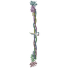
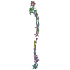
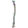
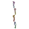
| Title | CRYSTAL STRUCTURE OF NATIVE CHICKEN FIBRINOGEN | ||||||
 Components Components | (FIBRINOGEN) x 3 | ||||||
 Keywords Keywords | BLOOD CLOTTING / coiled coils / disulfide rings / fibrin forming entities | ||||||
| Function / homology |  Function and homology information Function and homology informationblood coagulation, common pathway / fibrinogen complex / positive regulation of heterotypic cell-cell adhesion / blood coagulation, fibrin clot formation / protein polymerization / fibrinolysis / cell-matrix adhesion / platelet activation / platelet aggregation / : ...blood coagulation, common pathway / fibrinogen complex / positive regulation of heterotypic cell-cell adhesion / blood coagulation, fibrin clot formation / protein polymerization / fibrinolysis / cell-matrix adhesion / platelet activation / platelet aggregation / : / protein-macromolecule adaptor activity / signaling receptor binding / extracellular space / metal ion binding Similarity search - Function | ||||||
| Biological species |  | ||||||
| Method |  X-RAY DIFFRACTION / X-RAY DIFFRACTION /  SYNCHROTRON / Resolution: 5.5 Å SYNCHROTRON / Resolution: 5.5 Å | ||||||
 Authors Authors | Yang, Z. / Mochalkin, I. / Veerapandian, L. / Riley, M. / Doolittle, R.F. | ||||||
 Citation Citation |  Journal: Proc.Natl.Acad.Sci.USA / Year: 2000 Journal: Proc.Natl.Acad.Sci.USA / Year: 2000Title: Crystal structure of native chicken fibrinogen at 5.5-A resolution. Authors: Yang, Z. / Mochalkin, I. / Veerapandian, L. / Riley, M. / Doolittle, R.F. #1:  Journal: Nature / Year: 1997 Journal: Nature / Year: 1997Title: Crystal Structures of Fragment D from Human Fibrinogen and its Crosslinked Countepart from Fibrin Authors: Spraggon, G. / Everse, S.J. / Doolittle, R.F. #2:  Journal: Biochemistry / Year: 1998 Journal: Biochemistry / Year: 1998Title: Crystal Structure of Fragment Double-D from Human Fibrin with Two Different Bound Ligands Authors: Everse, S.J. / Spraggon, G. / Veerapandian, L. / Riley, M. / Doolittle, R.F. | ||||||
| History |
|
- Structure visualization
| Structure viewer | Molecule:  Molmil Molmil Jmol/JSmol Jmol/JSmol |
|---|
- Downloads & links
- Download
| PDBx/mmCIF format |  1ei3.cif.gz 1ei3.cif.gz | 84.6 KB | Display |  PDBx/mmCIF format PDBx/mmCIF format |
|---|---|---|---|---|
| PDB format |  pdb1ei3.ent.gz pdb1ei3.ent.gz | 43.5 KB | Display |  PDB format PDB format |
| PDBx/mmJSON format |  1ei3.json.gz 1ei3.json.gz | Tree view |  PDBx/mmJSON format PDBx/mmJSON format | |
| Others |  Other downloads Other downloads |
-Validation report
| Arichive directory |  https://data.pdbj.org/pub/pdb/validation_reports/ei/1ei3 https://data.pdbj.org/pub/pdb/validation_reports/ei/1ei3 ftp://data.pdbj.org/pub/pdb/validation_reports/ei/1ei3 ftp://data.pdbj.org/pub/pdb/validation_reports/ei/1ei3 | HTTPS FTP |
|---|
-Related structure data
| Related structure data | |
|---|---|
| Similar structure data |
- Links
Links
- Assembly
| Deposited unit | 
| ||||||||
|---|---|---|---|---|---|---|---|---|---|
| 1 |
| ||||||||
| Unit cell |
|
- Components
| #1: Protein | Mass: 54288.000 Da / Num. of mol.: 2 / Fragment: ALPHA CHAIN / Source method: isolated from a natural source / Source: (natural)  #2: Protein | Mass: 52874.277 Da / Num. of mol.: 2 / Fragment: BETA CHAIN / Source method: isolated from a natural source / Source: (natural)  #3: Protein | Mass: 47000.098 Da / Num. of mol.: 2 / Fragment: GAMMA CHAIN / Source method: isolated from a natural source / Source: (natural)  |
|---|
-Experimental details
-Experiment
| Experiment | Method:  X-RAY DIFFRACTION / Number of used crystals: 3 X-RAY DIFFRACTION / Number of used crystals: 3 |
|---|
- Sample preparation
| Crystal | Density Matthews: 3.81 Å3/Da / Density % sol: 67.75 % | ||||||||||||||||||||||||||||||||||||||||||||||||
|---|---|---|---|---|---|---|---|---|---|---|---|---|---|---|---|---|---|---|---|---|---|---|---|---|---|---|---|---|---|---|---|---|---|---|---|---|---|---|---|---|---|---|---|---|---|---|---|---|---|
| Crystal grow | Temperature: 298 K / Method: vapor diffusion, sitting drop / pH: 6.75 Details: PEG 3350, imidazole, calcium chloride, pH 6.75, VAPOR DIFFUSION, SITTING DROP, temperature 298K | ||||||||||||||||||||||||||||||||||||||||||||||||
| Crystal | *PLUS Density % sol: 65 % | ||||||||||||||||||||||||||||||||||||||||||||||||
| Crystal grow | *PLUS pH: 7 | ||||||||||||||||||||||||||||||||||||||||||||||||
| Components of the solutions | *PLUS
|
-Data collection
| Diffraction | Mean temperature: 298 K |
|---|---|
| Diffraction source | Source:  SYNCHROTRON / Site: SYNCHROTRON / Site:  NSLS NSLS  / Beamline: X12C / Wavelength: 1.04 / Beamline: X12C / Wavelength: 1.04 |
| Detector | Type: BRANDEIS / Detector: CCD / Date: Oct 29, 1999 |
| Radiation | Protocol: SINGLE WAVELENGTH / Monochromatic (M) / Laue (L): M / Scattering type: x-ray |
| Radiation wavelength | Wavelength: 1.04 Å / Relative weight: 1 |
| Reflection | Resolution: 5→50 Å / Num. all: 113182 / Num. obs: 45940 / % possible obs: 80 % / Observed criterion σ(F): 2 / Observed criterion σ(I): 2 / Redundancy: 2.8 % / Rmerge(I) obs: 0.09 / Net I/σ(I): 7.5 |
| Reflection | *PLUS Num. obs: 16412 / Num. measured all: 45940 |
| Reflection shell | *PLUS Highest resolution: 5.5 Å / Lowest resolution: 5.9 Å / % possible obs: 65.4 % |
- Processing
| Software |
| ||||||||||||
|---|---|---|---|---|---|---|---|---|---|---|---|---|---|
| Refinement | Highest resolution: 5.5 Å | ||||||||||||
| Refinement step | Cycle: LAST / Highest resolution: 5.5 Å
| ||||||||||||
| Refinement | *PLUS Highest resolution: 5.5 Å | ||||||||||||
| Solvent computation | *PLUS | ||||||||||||
| Displacement parameters | *PLUS |
 Movie
Movie Controller


 PDBj
PDBj


